AQA · A-Level · Biology
Cell Structure
This topic covers the fundamental building blocks of all living organisms: cells. You'll learn the differences between plant, animal, and bacterial cells, how they specialise to perform specific functions, and why stem cells hold such incredible medical potential.
- 6 min read
- 3 worked examples
- 5 practice questions
- 6 key terms
Study Notes
Overview
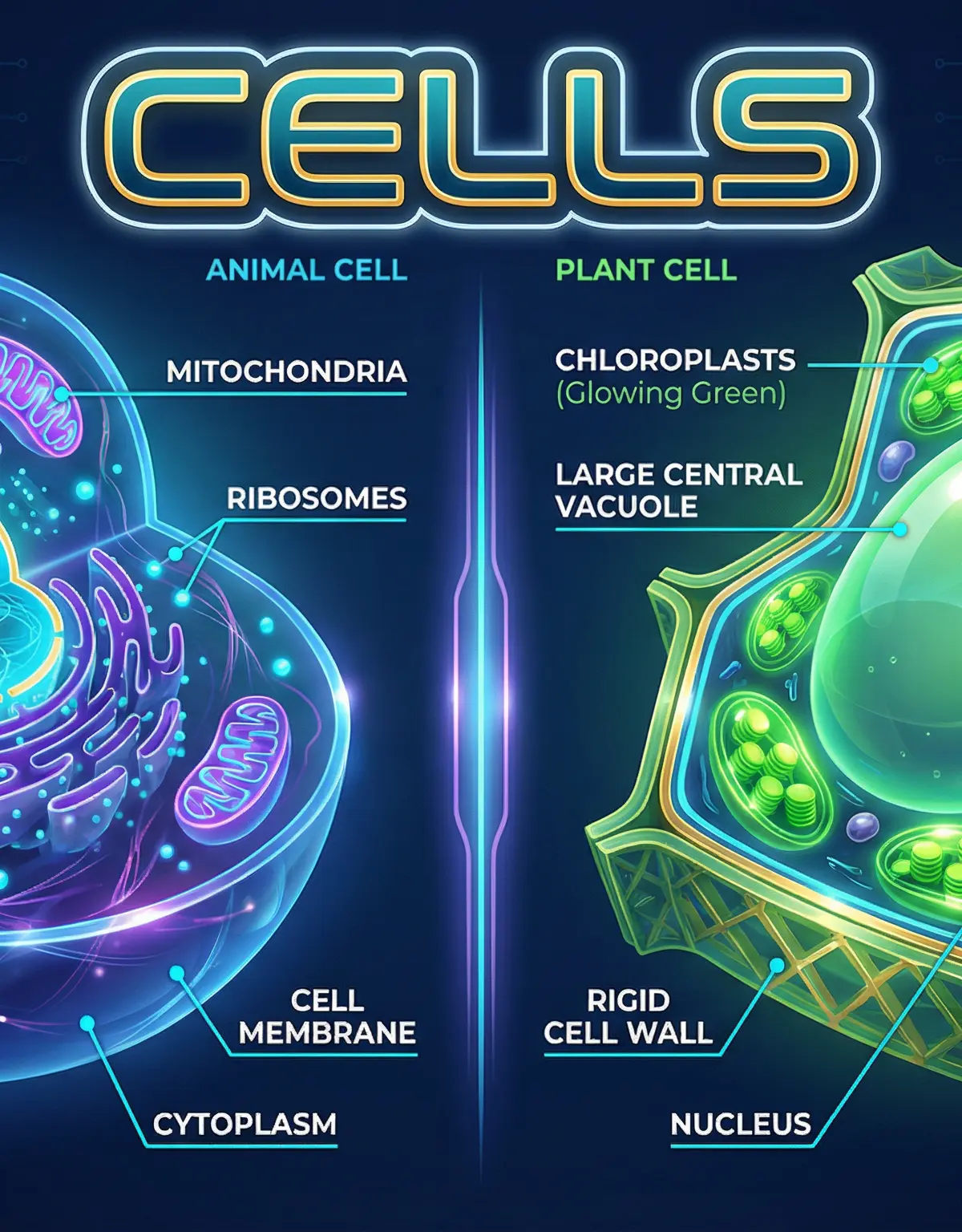
Cells are the basic structural and functional units of all living organisms. Whether you are studying human biology, plant physiology, or disease, everything begins at the cellular level. This topic is crucial because it forms the foundation for understanding how organisms grow, reproduce, and function. Examiners frequently test this topic, often asking you to compare different cell types, explain how organelles function, or describe how a cell's structure is adapted to its specific role. You will also explore cell differentiation and the controversial yet promising field of stem cell research.
Listen to the 10-minute podcast overview here:
Key Concepts
Concept 1: Eukaryotic vs Prokaryotic Cells
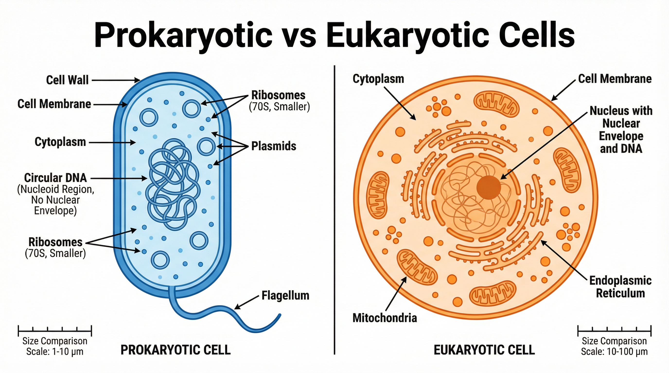
All cells can be divided into two main categories: eukaryotic and prokaryotic.
Eukaryotic cells are complex and include all animal and plant cells. Their defining feature is that their genetic material (DNA) is enclosed within a true nucleus. They are generally larger, ranging from 10 to 100 micrometres (μm).
Prokaryotic cells, such as bacteria, are much simpler and smaller, typically ranging from 1 to 10 μm. They do not have a nucleus. Instead, their genetic material is a single loop of DNA that floats freely in the cytoplasm, in a region called the nucleoid. Prokaryotes may also contain small rings of DNA called plasmids.
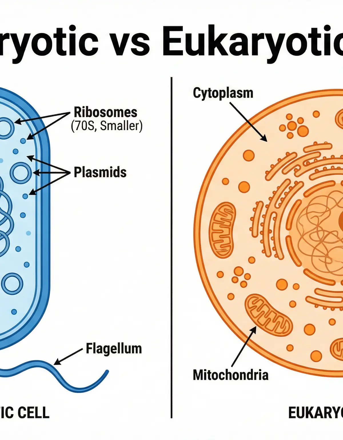
Example: If a bacterial cell is 2 μm long and an animal cell is 50 μm long, the animal cell is 25 times larger. Examiners often ask you to calculate magnification or actual size using the formula: Magnification = Image Size / Actual Size.
Concept 2: Animal and Plant Cells (Organelles)
Both animal and plant cells contain several key subcellular structures, known as organelles:
- Nucleus: Contains genetic material that controls the activities of the cell.
- Cytoplasm: A gel-like substance where most chemical reactions take place. It contains enzymes that control these reactions.
- Cell Membrane: Holds the cell together and controls what goes in and out (selectively permeable).
- Mitochondria: These are the site of most of the reactions for aerobic respiration. Respiration transfers energy that the cell needs to work.
- Ribosomes: These are where proteins are made in the cell (protein synthesis).
Plant cells have three additional structures that animal cells do not:
- Rigid Cell Wall: Made of cellulose. It supports the cell and strengthens it.
- Permanent Vacuole: Contains cell sap, a weak solution of sugar and salts. It helps maintain turgor pressure to keep the cell rigid.
- Chloroplasts: These are where photosynthesis occurs, which makes food for the plant. They contain a green substance called chlorophyll, which absorbs the light needed for photosynthesis.
Concept 3: Cell Differentiation and Specialisation
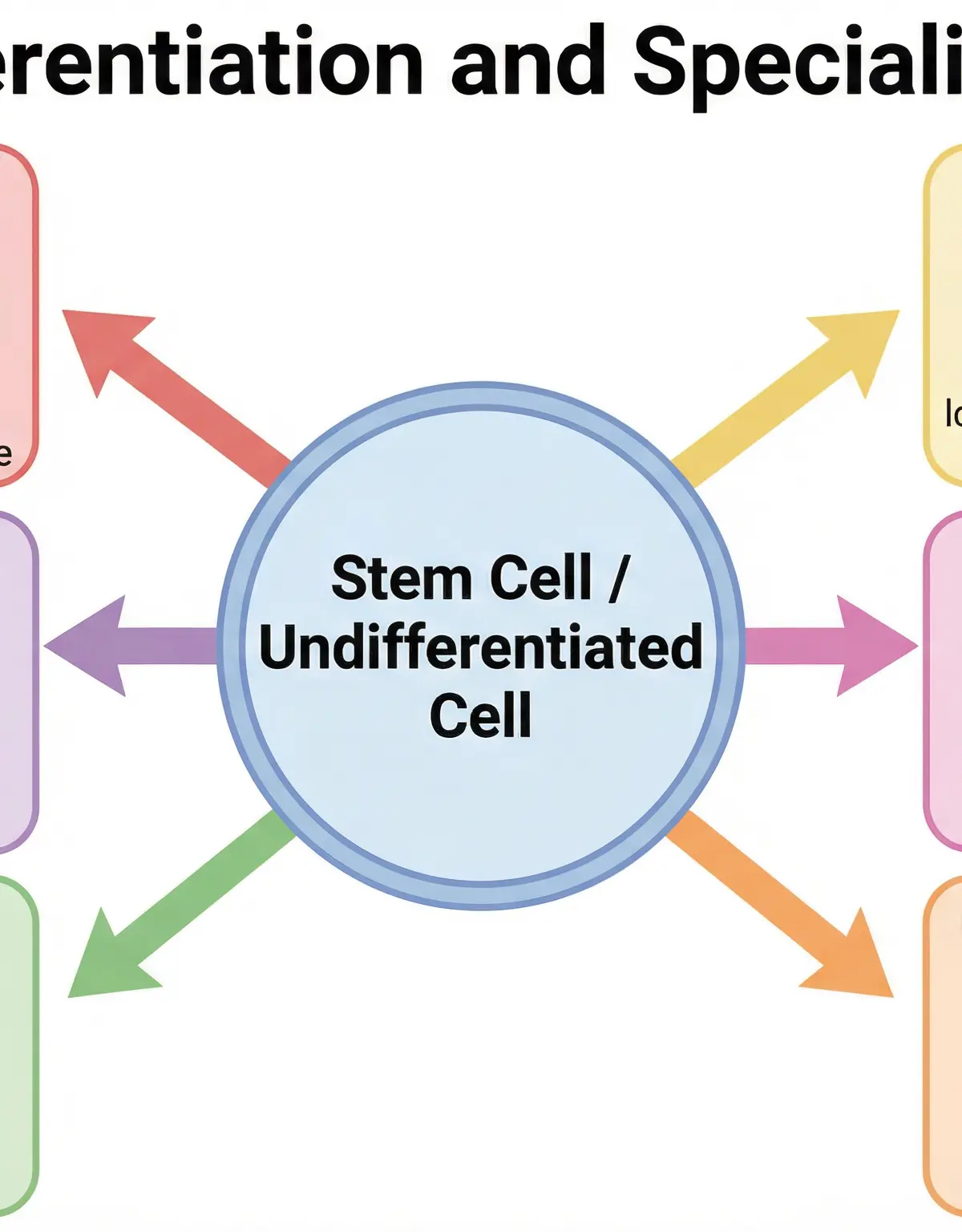
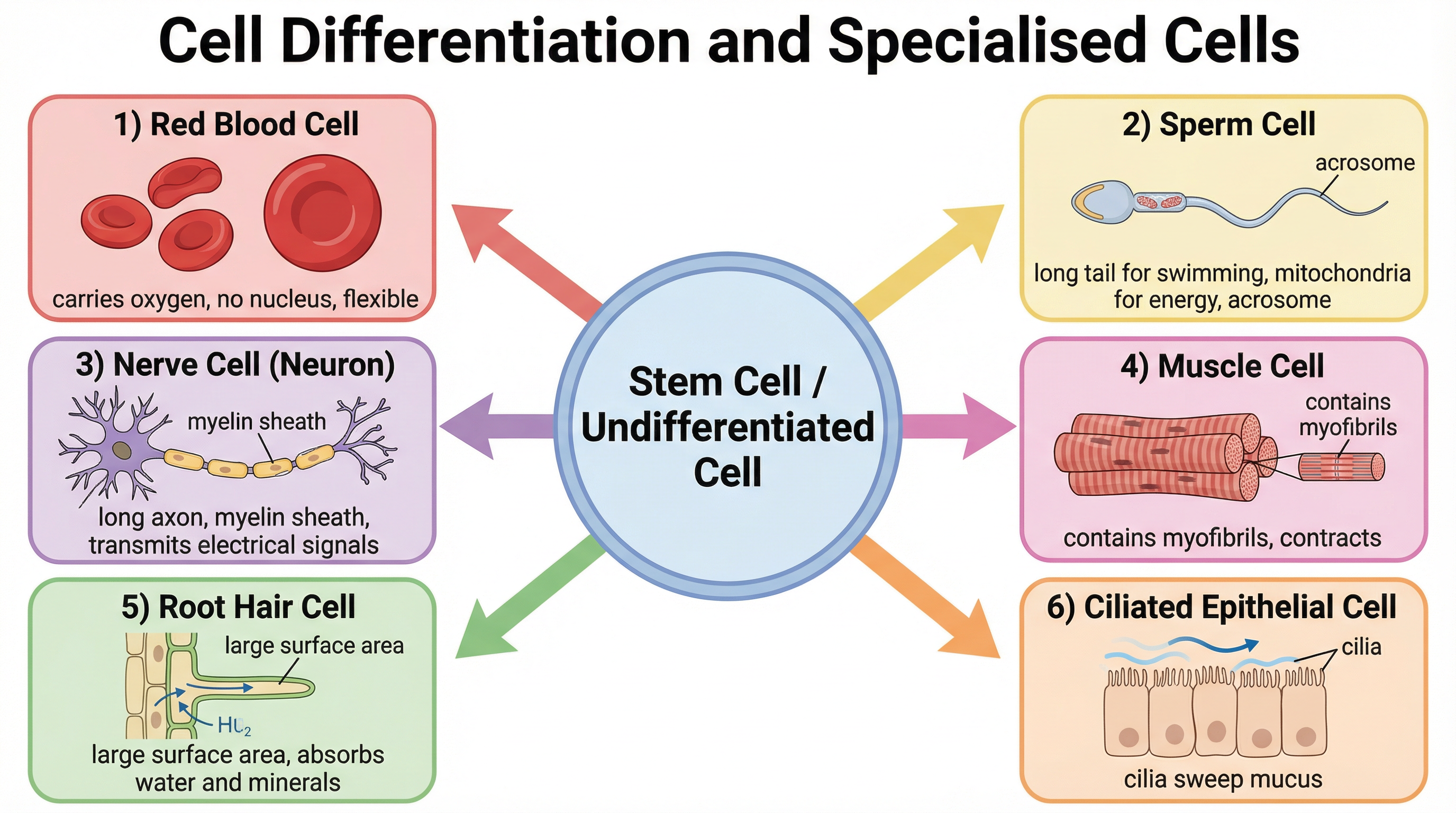
Differentiation is the process by which a cell changes to become specialised for its job. As cells change, they develop different subcellular structures and turn into different types of cells. This allows them to carry out specific functions.
In most animal cells, the ability to differentiate is lost at an early stage, after they become specialised. However, lots of plant cells don't ever lose this ability. The cells that differentiate in mature animals are mainly used for repairing and replacing cells, such as skin or blood cells.
Example: A sperm cell is specialised for reproduction. Its function is to get the male DNA to the female DNA. It has a long tail and a streamlined head to help it swim to the egg. There are lots of mitochondria in the cell to provide the energy needed. It also carries enzymes in its head (the acrosome) to digest through the egg cell membrane.
Concept 4: Stem Cells
Stem cells are undifferentiated cells that can divide to produce lots more undifferentiated cells. They can differentiate into different types of cells, depending on what instructions they're given.
Stem cells are found in early human embryos. These embryonic stem cells are incredibly versatile—they have the potential to turn into any kind of cell at all. Adults also have stem cells, but they're only found in certain places, like bone marrow. Unlike embryonic stem cells, adult stem cells can't turn into any cell type at all, only certain ones, such as blood cells.
Stem cells from embryos and bone marrow can be grown in a lab to produce clones (genetically identical cells) and made to differentiate into specialised cells to use in medicine or research. However, there are ethical issues surrounding the use of embryonic stem cells, as some people feel that human embryos shouldn't be used for experiments since each one is a potential human life.
Mathematical/Scientific Relationships
Magnification Equation:
Magnification = Image Size / Actual Size
(You must memorise this formula. It is not given on the formula sheet.)
Unit Conversions:
Examiners frequently test your ability to convert between units of length.
- 1 millimetre (mm) = 1000 micrometres (μm)
- 1 micrometre (μm) = 1000 nanometres (nm)
Tip: To convert mm to μm, multiply by 1000. To convert μm to mm, divide by 1000.
Practical Applications
Required Practical: Using a Light MicroscopeExaminers will test your knowledge of how to prepare a slide and use a microscope.
- Method: To look at onion cells, add a drop of water to the middle of a clean slide. Cut up an onion and separate it out into layers. Use tweezers to peel off some epidermal tissue from the bottom of one of the layers. Place the epidermal tissue into the water on the slide. Add a drop of iodine solution. Iodine is a stain—it highlights objects in a cell by adding colour to them. Place a cover slip on top.
- Using the Microscope: Clip the slide onto the stage. Select the lowest-powered objective lens. Use the coarse adjustment knob to move the stage up to just below the objective lens. Look down the eyepiece. Use the coarse adjustment knob to move the stage downwards until the image is roughly in focus. Adjust the focus with the fine adjustment knob, until you get a clear image of what's on the slide.
- Common Errors: Getting air bubbles under the cover slip (which look like large, thick-rimmed circles), or not using the lowest power objective lens first.
Visual Resources
2 diagrams and illustrations
Interactive Diagrams
2 interactive diagrams to visualise key concepts
Conceptual Flow Outline
Flowchart showing how an undifferentiated stem cell can differentiate into various specialised cells with specific functions.
Conceptual Flow Outline
Simple process diagram showing how a light microscope magnifies an image.
Worked Examples
3 worked examples — open one to explore the question and available guidance.
Practice Questions
Test your understanding — click to reveal model answers
State the function of ribosomes. (1 mark)
Hint: Think about what is being synthesised or 'made' here.
A student views a cheek cell under a light microscope. The eyepiece lens is x10 and the objective lens is x40. Calculate the total magnification. (1 mark)
Hint: How do you combine the magnification of two lenses?
Explain how an electron microscope has helped our understanding of cells compared to a light microscope. (3 marks)
Hint: Think about the two key terms: magnification and resolution.
Evaluate the use of embryonic stem cells compared to adult bone marrow stem cells in medical treatments. (6 marks)
Hint: Evaluate means you need pros and cons for BOTH, and a conclusion.
A bacterial cell has a length of 2.5 μm. Calculate its length in millimetres. Give your answer in standard form. (2 marks)
Hint: First convert μm to mm, then put that number into standard form.