Subject: Biology | Level: GCSE | Exam Board: WJEC
Master the foundational building blocks of biology: prokaryotic and eukaryotic cells. This guide covers everything from sub-cellular structures and their functions to the development of microscopy, ensuring you can confidently tackle diagrams, comparisons, and magnification calculations.
Revision Notes & Key Concepts
Revision Podcast Transcript
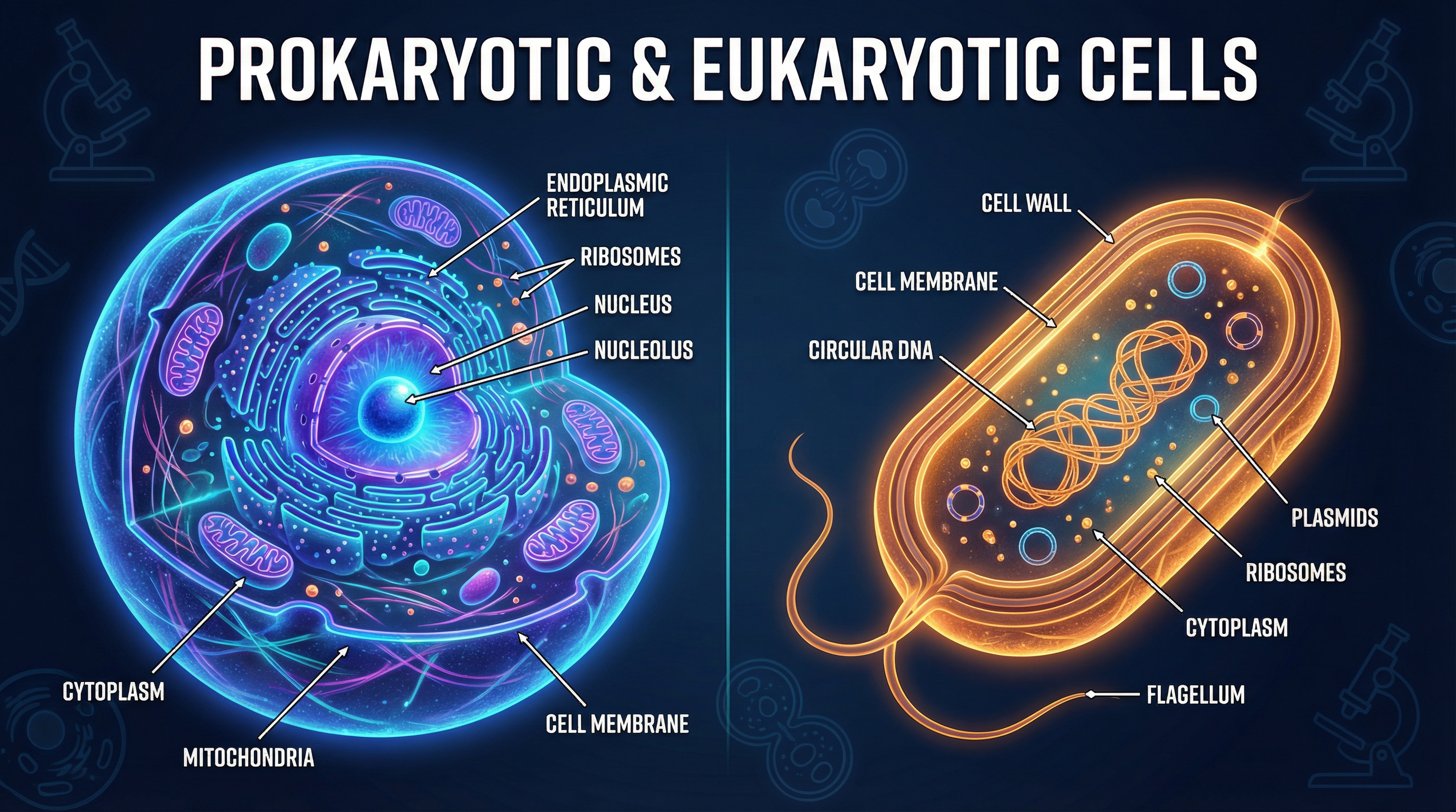
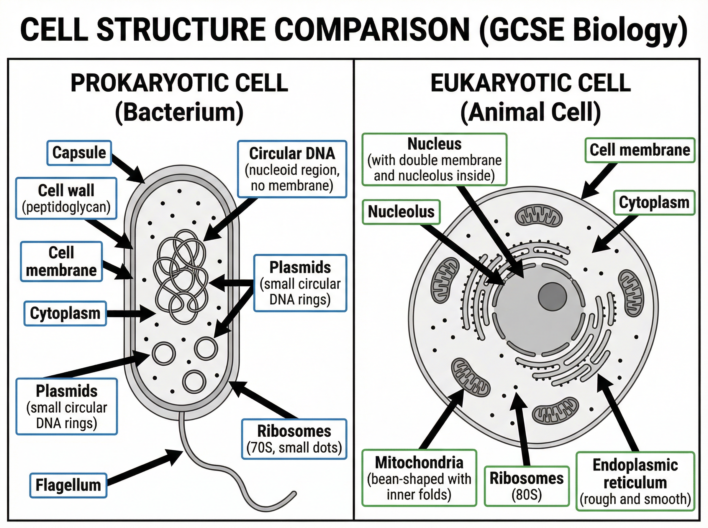
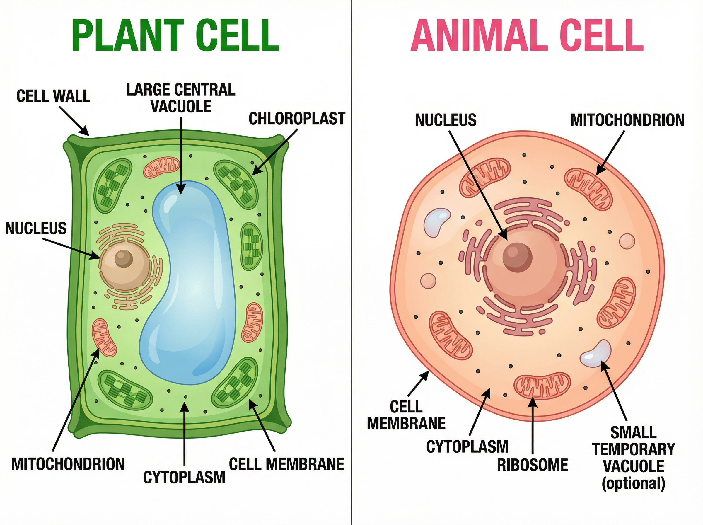
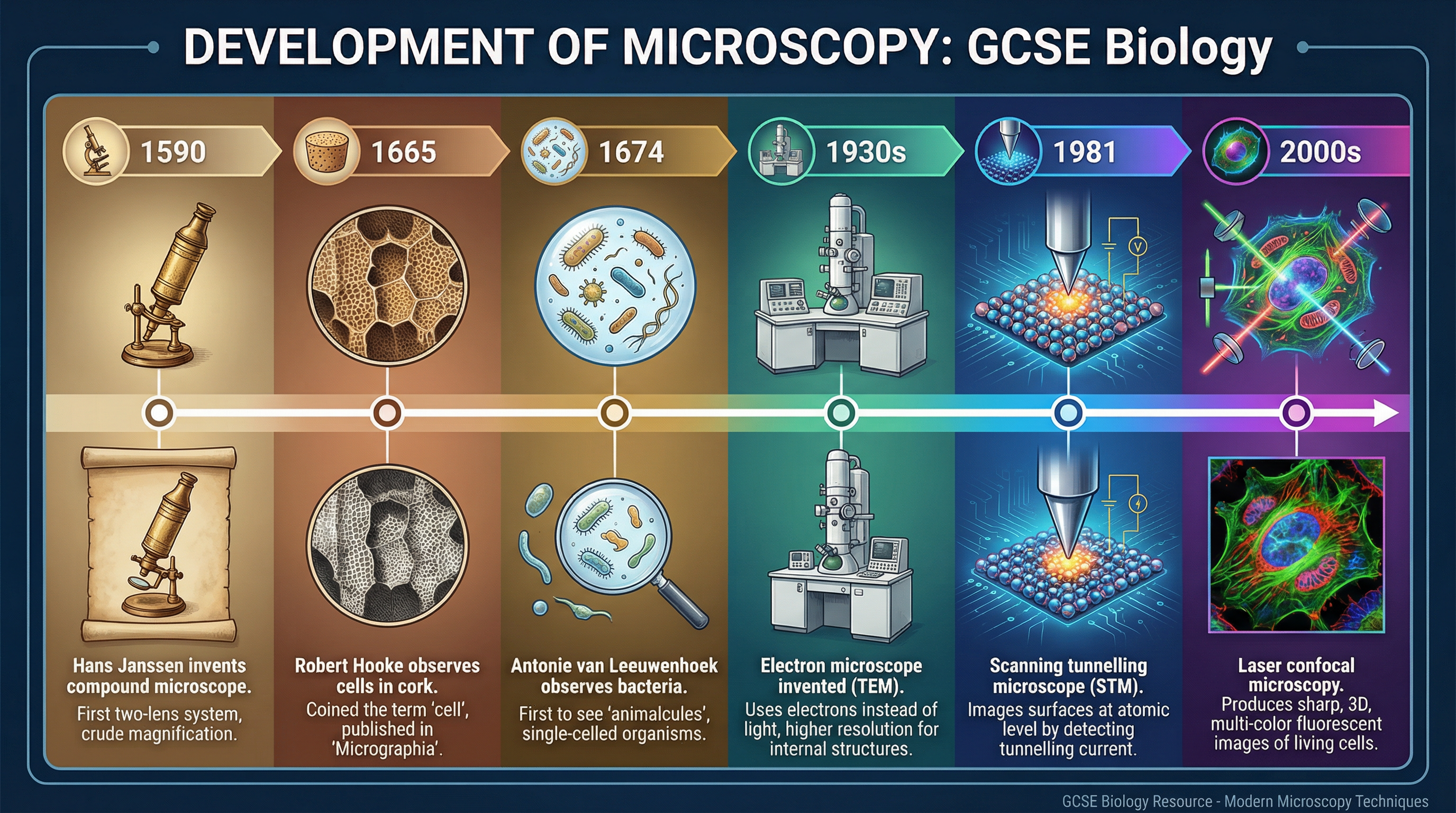
Welcome to your GCSE Biology revision podcast. I'm your tutor today, and we're diving into one of the most fundamental topics in the entire specification: Prokaryotic and Eukaryotic Cells — that's topic 1.1. Whether you're revising for AQA, Edexcel, or OCR, this topic comes up every single year, so let's make sure you absolutely nail it. By the end of this episode, you'll be able to describe the key differences between prokaryotic and eukaryotic cells, explain the function of every sub-cellular structure you need to know, draw and label cell diagrams accurately, and tackle any exam question on microscopy with confidence. Let's get started. SECTION ONE: THE BIG PICTURE — WHAT ARE CELLS? All living things are made of cells. That's the starting point. But not all cells are the same. In biology, we divide all cells into two fundamental categories: prokaryotic cells and eukaryotic cells. The word "prokaryotic" comes from the Greek meaning "before nucleus." These are the simpler, smaller, more ancient cells — bacteria are the classic example. Eukaryotic cells, on the other hand, have a true nucleus. The word means "true nucleus" in Greek. Animals, plants, fungi, and protists are all made of eukaryotic cells. Think of it this way: a prokaryotic cell is like a studio flat — everything is in one open space, no separate rooms. A eukaryotic cell is like a house with multiple rooms — each organelle has its own compartment, separated by membranes. This distinction is absolutely fundamental. Examiners will test it directly, so you need to be crystal clear on which structures are found in which type of cell. SECTION TWO: PROKARYOTIC CELLS IN DETAIL Let's start with prokaryotic cells, using a bacterium as our model. A typical bacterial cell is between one and ten micrometres in length — that's tiny. For context, a human hair is about 70 micrometres wide. Here are the structures you must know for a prokaryotic cell: First, the cell wall. Bacteria have a cell wall made of a substance called peptidoglycan. This is different from plant cell walls, which are made of cellulose. The cell wall gives the bacterium its shape and prevents it from bursting due to osmosis. This is an important distinction — examiners often ask candidates to compare cell walls in different cell types. Second, the cell membrane. Just inside the cell wall, there's a cell membrane made of a phospholipid bilayer. This controls what enters and leaves the cell. It's selectively permeable — meaning it lets some substances through but not others. Third, the cytoplasm. This is the jelly-like fluid that fills the cell. All the chemical reactions of life take place here. It contains enzymes, salts, and other molecules needed for metabolism. Fourth, ribosomes. These are tiny structures found throughout the cytoplasm. Their job is to make proteins — a process called protein synthesis. Bacterial ribosomes are slightly smaller than eukaryotic ribosomes. Bacterial ribosomes are 70S in size, while eukaryotic ribosomes are 80S. This difference is actually exploited by antibiotics — many antibiotics work by targeting bacterial ribosomes without affecting our own. Fifth, and this is crucial — the genetic material. In prokaryotic cells, the DNA is not enclosed in a nucleus. Instead, it floats freely in the cytoplasm as a single circular chromosome. This is sometimes called the nucleoid region. Examiners love to test this — candidates frequently lose marks by saying bacteria have a nucleus. They do not. Sixth, plasmids. These are small, circular loops of DNA found in the cytoplasm, separate from the main circular chromosome. Plasmids carry additional genes — often ones that give the bacterium an advantage, like antibiotic resistance. Not all bacteria have plasmids, but you must know what they are and where they're found. Some bacteria also have additional structures: a flagellum — that's a long, whip-like tail used for movement. And some have a capsule — a slimy outer layer that helps the bacterium stick to surfaces and evade the immune system. So to summarise a prokaryotic cell: cell wall, cell membrane, cytoplasm, ribosomes, circular DNA in the cytoplasm, and plasmids. No nucleus, no mitochondria, no chloroplasts. SECTION THREE: EUKARYOTIC CELLS IN DETAIL Now let's look at eukaryotic cells. We'll cover animal cells first, then plant cells, because plants have some extra structures. ANIMAL CELLS The nucleus is the control centre of the cell. It contains the cell's DNA in the form of chromosomes. The nucleus is surrounded by a double membrane called the nuclear envelope, which has pores in it to allow molecules in and out. Inside the nucleus, you'll find the nucleolus — a darker region where ribosomes are made. The mitochondria — singular: mitochondrion — are the powerhouses of the cell. This is where aerobic respiration takes place, releasing energy in the form of ATP. Mitochondria have a double membrane — the inner membrane is folded into structures called cristae, which increase the surface area for respiration reactions. Cells that need lots of energy — like muscle cells — have many mitochondria. The cell membrane controls what enters and leaves the cell, just like in prokaryotic cells. It's made of a phospholipid bilayer with proteins embedded in it. The cytoplasm is the fluid medium where most cellular reactions occur. Ribosomes in eukaryotic cells are slightly larger than in prokaryotic cells — 80S — but their function is the same: protein synthesis. PLANT CELLS Plant cells have everything animal cells have, plus three extra structures: The cell wall. Plant cell walls are made of cellulose — a strong, rigid material. The cell wall gives the plant cell its shape and provides structural support. Remember: bacterial cell walls are made of peptidoglycan, plant cell walls are made of cellulose. Examiners will test this distinction. Chloroplasts. These are the organelles where photosynthesis takes place. They contain a green pigment called chlorophyll, which absorbs light energy. Chloroplasts have a double membrane and contain stacks of membranes called thylakoids, arranged into structures called grana. Only plant cells and algae have chloroplasts — animal cells do not. The permanent vacuole. This is a large, fluid-filled sac in the centre of a plant cell. It's filled with cell sap — a solution of sugars, salts, and other substances. The vacuole helps maintain the cell's shape by pushing outwards against the cell wall, creating turgor pressure. Animal cells may have small, temporary vacuoles, but not the large permanent one found in plant cells. SECTION FOUR: KEY COMPARISON TABLE Let me give you a quick comparison to lock this in. Prokaryotic cells: no nucleus, circular DNA free in cytoplasm, plasmids present, 70S ribosomes, cell wall made of peptidoglycan, no mitochondria, no chloroplasts. Size: 1 to 10 micrometres. Eukaryotic animal cells: nucleus present with linear DNA, no plasmids, 80S ribosomes, cell membrane only — no cell wall, mitochondria present, no chloroplasts. Size: 10 to 100 micrometres. Eukaryotic plant cells: nucleus present, no plasmids, 80S ribosomes, cell wall made of cellulose, mitochondria present, chloroplasts present, large permanent vacuole. Size: 10 to 100 micrometres. SECTION FIVE: MICROSCOPY Now let's talk about microscopes — because you need to understand how scientists actually see these cells, and how microscopy technology has developed over time. THE LIGHT MICROSCOPE The light microscope, also called the optical microscope, uses visible light and lenses to magnify specimens. It can magnify up to about 1,500 times and has a resolution of about 200 nanometres. Resolution is the ability to distinguish between two points as separate — the higher the resolution, the more detail you can see. Light microscopes are great for viewing living cells and tissues. You can watch cells dividing in real time. However, they cannot show the fine details of sub-cellular structures like ribosomes or the internal structure of mitochondria. THE ELECTRON MICROSCOPE The electron microscope, developed in the 1930s, uses beams of electrons instead of light. Because electrons have a much shorter wavelength than light, electron microscopes have a much higher resolution — down to 0.1 nanometres. This means they can magnify up to 500,000 times or more. There are two main types. The Transmission Electron Microscope, or TEM, passes electrons through a thin slice of the specimen. It produces detailed images of internal structures — this is how we first saw the internal structure of mitochondria and the double membrane of the nucleus. The Scanning Electron Microscope, or SEM, bounces electrons off the surface of a specimen, producing three-dimensional images of surfaces. The downside of electron microscopes? Specimens must be dead and specially prepared — you can't observe living processes. They're also very expensive and large. LASER CONFOCAL MICROSCOPY More recently, laser confocal microscopes use lasers and fluorescent dyes to produce sharp, three-dimensional images of living cells. This has revolutionised our understanding of how organelles move and interact in real time. MAGNIFICATION FORMULA You need to know this formula: Magnification equals image size divided by actual size. Or written as a formula: M equals I divided by A. So if an image of a cell is 5 centimetres wide and the actual cell is 50 micrometres wide, the magnification is 5 centimetres divided by 50 micrometres. But you must use the same units! Convert 5 centimetres to 50,000 micrometres. So magnification equals 50,000 divided by 50, which equals 1,000 times magnification. Always show your working and include the multiplication sign and the letter x after the number. SECTION SIX: EXAM TIPS AND COMMON MISTAKES Right, let's talk exam technique. These are the mistakes I see candidates make time and time again. MISTAKE ONE: Saying bacteria have a nucleus. They do not. Bacteria have circular DNA free in the cytoplasm. Never write "nucleus" for a prokaryotic cell. You will lose the mark every time. MISTAKE TWO: Confusing mitochondria and chloroplasts. Mitochondria are for respiration — releasing energy. Chloroplasts are for photosynthesis — making food using light. A common wrong answer is saying chloroplasts release energy. They don't — they capture it. Mitochondria release it. MISTAKE THREE: Forgetting plasmids. When asked to describe the genetic material in a prokaryotic cell, many candidates only mention the circular chromosome and forget to mention plasmids. Always mention both. MISTAKE FOUR: Getting the cell wall material wrong. Plant cell walls are cellulose. Bacterial cell walls are peptidoglycan. If a question asks you to compare them, you must name the specific material. MISTAKE FIVE: Diagram labelling errors. When drawing a plant cell, you must include the cell wall, large vacuole, and chloroplasts. When drawing an animal cell, you must NOT include these. Examiners will penalise you for including structures that shouldn't be there. EXAM TECHNIQUE FOR DESCRIBE AND EXPLAIN QUESTIONS For a "describe" question — just say what you see or what happens. Use correct terminology. For example: "The nucleus contains the cell's DNA." For an "explain" question — you must say WHY or HOW. Use the word "because" or "so that" to link cause and effect. For example: "Mitochondria have folded inner membranes because this increases the surface area for the reactions of aerobic respiration, allowing more ATP to be produced." For a "compare" question — you must address BOTH things being compared. Don't just describe one. Use comparative language: "whereas," "in contrast," "unlike." For example: "Prokaryotic cells have circular DNA free in the cytoplasm, whereas eukaryotic cells have linear DNA enclosed within a membrane-bound nucleus." For a six-mark question — structure your answer clearly. Cover at least six distinct points. Don't repeat yourself. Use paragraphs or a logical sequence. SECTION SEVEN: QUICK-FIRE RECALL QUIZ Okay, let's test what you've learned. I'll ask a question — pause the podcast, think of your answer, then I'll give you the correct response. Question one: Name THREE structures found in a prokaryotic cell but NOT in a eukaryotic animal cell. Answer: Plasmids, cell wall made of peptidoglycan, and circular DNA free in the cytoplasm. Flagellum is also acceptable. Question two: What is the function of the mitochondria? Answer: The site of aerobic respiration, where glucose and oxygen are used to release energy in the form of ATP. Question three: What is the difference in resolution between a light microscope and an electron microscope? Answer: A light microscope has a resolution of about 200 nanometres. An electron microscope has a resolution of about 0.1 nanometres — so it can show much finer detail. Question four: A cell image measures 4 centimetres. The actual cell is 20 micrometres. What is the magnification? Answer: Convert 4 centimetres to 40,000 micrometres. Magnification equals 40,000 divided by 20, which equals 2,000 times. Write it as times 2000. Question five: Name TWO structures found in plant cells but NOT in animal cells. Answer: Cell wall made of cellulose, chloroplasts, and permanent vacuole. Any two of these would earn the marks. SECTION EIGHT: SUMMARY AND SIGN-OFF Let's bring it all together. Here are the five most important things to take away from this episode. One: Prokaryotic cells have no nucleus — their circular DNA floats freely in the cytoplasm. Eukaryotic cells have a membrane-bound nucleus. Two: Prokaryotic cells also have plasmids — small circular loops of extra DNA. Don't forget to mention these. Three: Plant cells have three structures animal cells don't: a cell wall made of cellulose, chloroplasts for photosynthesis, and a large permanent vacuole. Four: Electron microscopes have much higher resolution than light microscopes — they can show sub-cellular structures in detail. The magnification formula is M equals I over A. Five: In the exam, always use precise language. "Mitochondria are the site of aerobic respiration" — not just "they make energy." "The nucleus contains DNA in the form of chromosomes" — not just "it controls the cell." You've got this. Practice drawing and labelling those cell diagrams from memory — that's the single best thing you can do for this topic. And remember: every mark counts. Good luck with your revision, and I'll see you in the next episode.
Key Terms & Definitions
- Prokaryotic Cell
- A type of cell lacking a nucleus and membrane-bound organelles; its genetic material is a single loop of DNA free in the cytoplasm.
- Eukaryotic Cell
- A type of cell with a membrane-bound nucleus containing its genetic material, and other membrane-bound organelles.
- Plasmid
- A small, circular ring of DNA found in the cytoplasm of bacterial cells, separate from the main chromosomal DNA.
- Resolution
- The ability to distinguish between two separate points; the minimum distance apart that two objects can be in order for them to appear as separate items.
- Mitochondria
- The site of aerobic respiration in eukaryotic cells, where energy is released.
- Ribosome
- The sub-cellular structure responsible for protein synthesis.
Worked Examples
Worked Example
Question: A student observed a palisade mesophyll cell under a light microscope. The actual length of the cell was 50 µm. The student drew the cell, and the length of the drawing was 100 mm. Calculate the magnification of the drawing. [3 marks]
Solution: Step 1: Convert units so they are the same. Convert mm to µm. 100 mm x 1000 = 100,000 µm Step 2: State the formula. Magnification = Image Size / Actual Size Step 3: Substitute the values and calculate. Magnification = 100,000 / 50 Final answer: x2000
Worked Example
Question: Compare the structure of a typical bacterial cell with a typical plant cell. [6 marks]
Solution: Both a bacterial cell and a plant cell have a cell membrane, cytoplasm, and ribosomes. They also both have a cell wall, but the bacterial cell wall is made of peptidoglycan, whereas the plant cell wall is made of cellulose. However, there are several key differences. A plant cell is eukaryotic, so its genetic material is enclosed within a nucleus. In contrast, a bacterial cell is prokaryotic, so its genetic material is a single loop of DNA free in the cytoplasm. Furthermore, bacterial cells may contain small rings of DNA called plasmids, which plant cells do not have. Finally, plant cells contain mitochondria and chloroplasts, as well as a large permanent vacuole, whereas bacterial cells do not contain any of these membrane-bound organelles.
Worked Example
Question: Explain why an electron microscope is more suitable than a light microscope for studying the internal structure of a mitochondrion. [2 marks]
Solution: An electron microscope has a much higher resolving power (resolution) than a light microscope [1 mark]. This means it can distinguish between closer points, allowing finer details and smaller sub-cellular structures to be seen clearly [1 mark].
Practice Questions
Question: Name the substance that makes up the plant cell wall. [1 mark]
Answer:
Question: Describe the function of ribosomes and state where they are found in a cell. [2 marks]
Answer:
Question: A bacterial cell has a length of 2 µm. Calculate its length in millimetres. Give your answer in standard form. [2 marks]
Answer:
Question: A student claims that all cells have a nucleus. Evaluate this claim using examples. [4 marks]
Answer:
Question: Explain how the development of the electron microscope has increased our understanding of sub-cellular structures. [3 marks]
Answer: