Subject: Biology | Level: GCSE | Exam Board: WJEC
Master the human transport system! This guide covers the double circulatory system, heart structure, blood vessels, and blood components. Essential for securing marks on structure-function relationships.
Revision Notes & Key Concepts
Revision Podcast Transcript
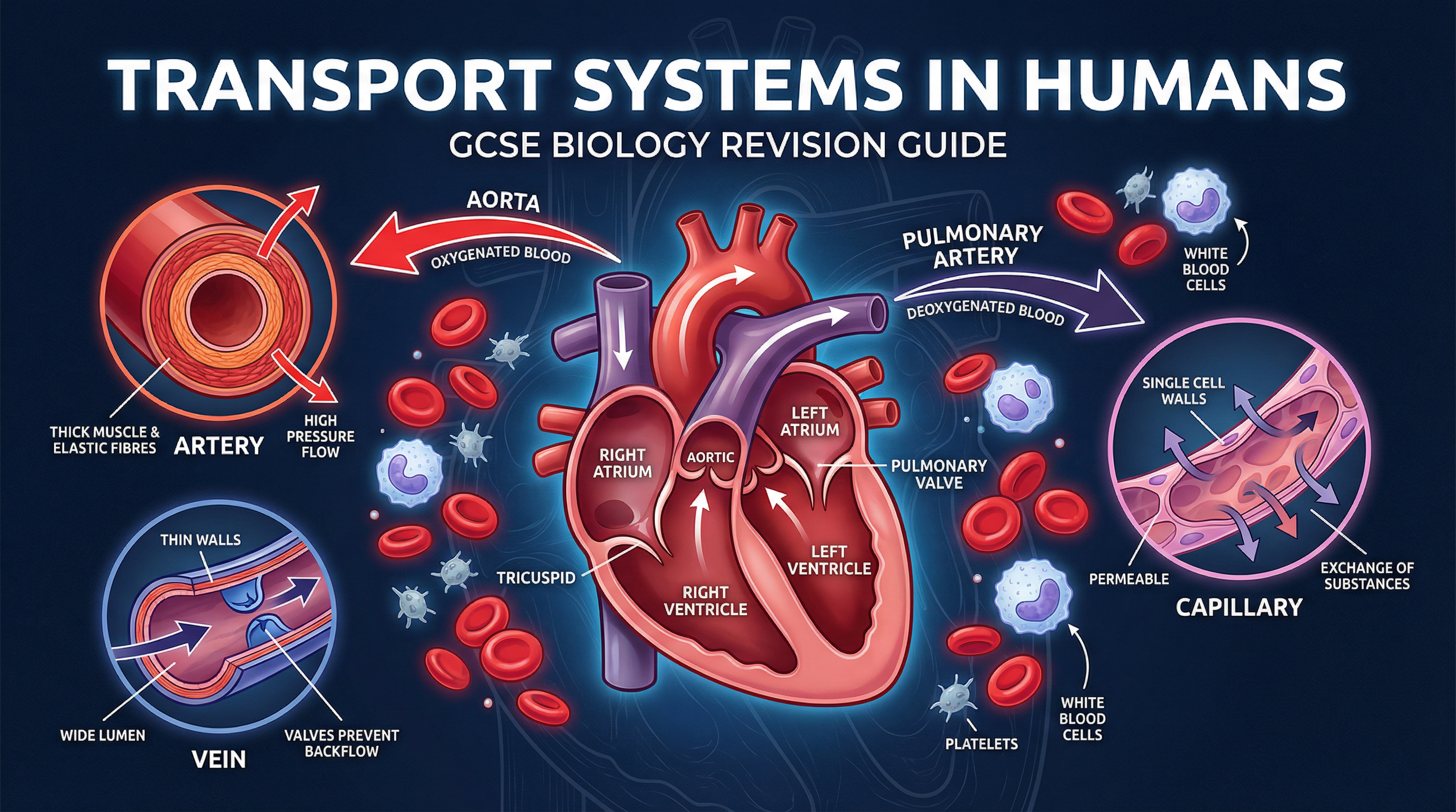
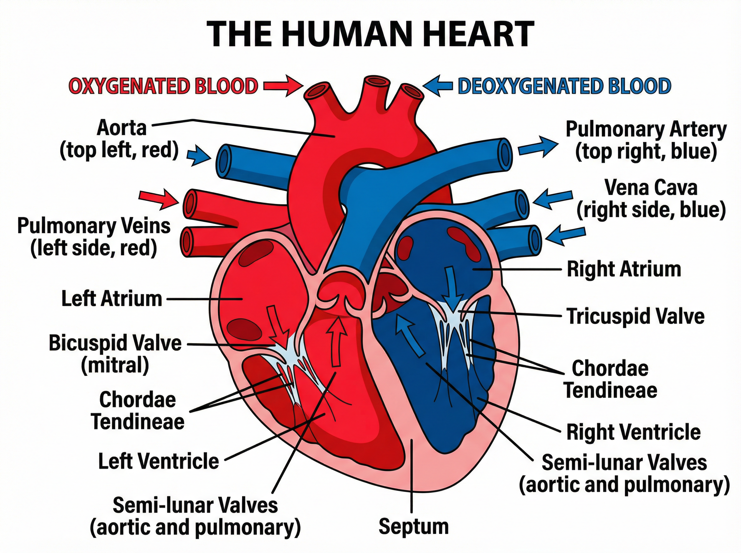
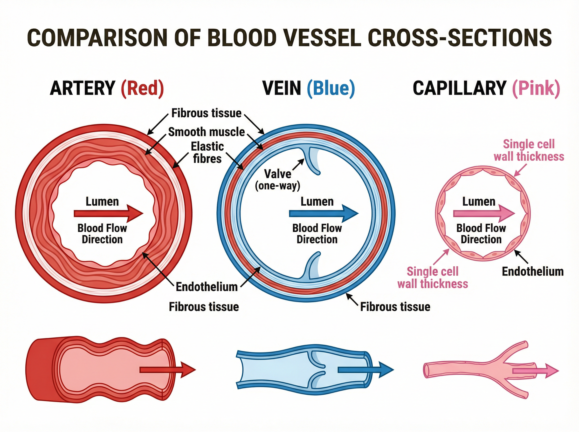
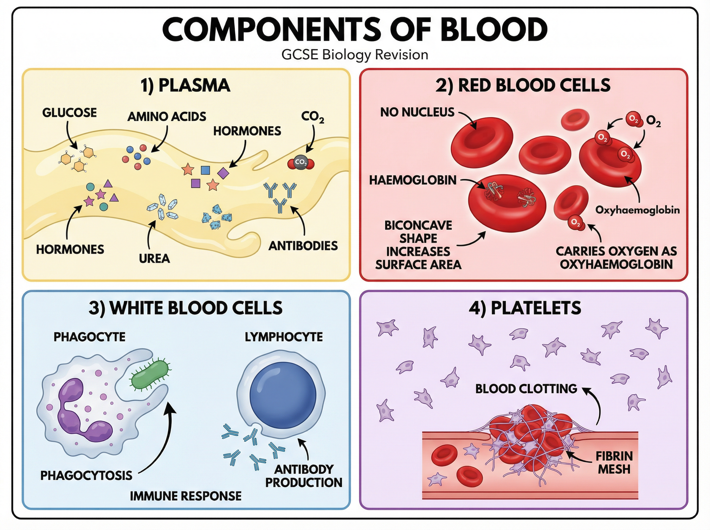
TRANSPORT SYSTEMS IN HUMANS — GCSE BIOLOGY REVISION PODCAST A 10-Minute Revision Episode [INTRO — approximately 1 minute] Hello and welcome! I'm so glad you've tuned in to this revision session. Today we're diving into one of the most fascinating and exam-rich topics in GCSE Biology — Transport Systems in Humans. Whether you're revising for the first time or doing a final polish before your exam, this episode is going to give you everything you need to feel confident and pick up those marks. By the end of this podcast, you'll be able to describe the double circulatory system, explain the structure and function of the heart, compare the three types of blood vessels, and describe the components of blood and what they do. These are all real exam favourites, and I'll be pointing out exactly where the marks are hiding throughout. So grab a pen and paper, and let's get started. [CORE CONCEPTS — approximately 5 minutes] Let's begin with the big picture: the double circulatory system. The human body uses what we call a double circulatory system. This means that blood passes through the heart twice in one complete circuit around the body. The first circuit is the pulmonary circulation — blood travels from the right side of the heart, to the lungs to pick up oxygen, and then back to the left side of the heart. The second circuit is the systemic circulation — blood travels from the left side of the heart, out to the rest of the body to deliver oxygen and nutrients, and then back to the right side of the heart. Why is this double system so important? Because it means oxygenated blood reaches the body at high pressure, making delivery of oxygen to cells much more efficient. Examiners love asking you to explain the advantage of a double circulatory system — always link it to maintaining high blood pressure for efficient delivery to tissues. Now let's talk about the heart itself. Think of the heart as a double pump — two pumps sitting side by side, separated by a muscular wall called the septum. The right side pumps deoxygenated blood to the lungs. The left side pumps oxygenated blood to the body. Each side has two chambers. The upper chambers are called atria — that's the plural of atrium. The lower chambers are the ventricles. So we have: right atrium, right ventricle, left atrium, and left ventricle. Blood flows in through the atria, which contract to push blood down into the ventricles, and then the ventricles contract powerfully to push blood out of the heart. Here's an important adaptation point that examiners test regularly: the left ventricle has a much thicker muscular wall than the right ventricle. Why? Because it needs to generate enough pressure to pump blood all the way around the entire body, whereas the right ventricle only needs to pump blood the short distance to the lungs. Thicker wall equals more muscle equals more force. Make sure you can explain this — it's worth marks. Now, the major blood vessels. Blood enters the right atrium from the body via the vena cava — that's the large vein returning deoxygenated blood. From the right ventricle, the pulmonary artery carries deoxygenated blood to the lungs. This is a classic exam trap — the pulmonary artery carries deoxygenated blood, even though arteries usually carry oxygenated blood. Don't fall for it! After picking up oxygen in the lungs, the pulmonary veins carry oxygenated blood back to the left atrium. Again — the pulmonary veins carry oxygenated blood, even though veins usually carry deoxygenated blood. From the left ventricle, the aorta — the largest artery in the body — carries oxygenated blood to the rest of the body. Let's talk about valves. The heart has four main valves, and their job is to prevent backflow of blood — to make sure blood only flows in one direction. Between the right atrium and right ventricle is the tricuspid valve. Between the left atrium and left ventricle is the bicuspid valve, also called the mitral valve. At the exit of each ventricle are the semi-lunar valves — the pulmonary semi-lunar valve on the right, and the aortic semi-lunar valve on the left. When a ventricle contracts, pressure builds up, the valve opens, blood flows out. When the ventricle relaxes, pressure drops, blood would flow back, but the valve snaps shut. That's the "lub-dub" sound of your heartbeat — valves closing. Now let's move on to blood vessels. There are three types: arteries, veins, and capillaries. Arteries carry blood away from the heart. They carry blood at high pressure, so they need thick, muscular, elastic walls to withstand and smooth out the pressure. They have a relatively narrow lumen — that's the central channel — and no valves, because the pressure keeps blood moving in the right direction. Veins carry blood back to the heart. Blood pressure is much lower in veins, so they have thinner walls and a wider lumen. Crucially, veins have valves to prevent backflow, because the pressure alone isn't enough to keep blood moving in one direction. Capillaries are the smallest blood vessels — so narrow that red blood cells have to pass through in single file. Their walls are just one cell thick, which gives a very short diffusion distance for substances to move between the blood and the surrounding cells. They form extensive networks — capillary beds — to ensure every cell in the body is close to a blood supply. This is where the actual exchange of oxygen, carbon dioxide, glucose, and waste products happens. Finally, let's cover the components of blood. Blood is made up of four main components. First, plasma — the liquid part of blood. It's a pale yellow fluid that transports dissolved substances including glucose, amino acids, hormones, carbon dioxide, urea, and antibodies. Plasma is the delivery and waste-collection service. Second, red blood cells — also called erythrocytes. They carry oxygen from the lungs to the body's cells. They're adapted in several brilliant ways: they have a biconcave disc shape, which increases their surface area for absorbing oxygen. They're packed with haemoglobin — the protein that binds to oxygen to form oxyhaemoglobin. And they have no nucleus, which means more space for haemoglobin. In the lungs, haemoglobin picks up oxygen. In the tissues, it releases it. Third, white blood cells — also called leucocytes. They're part of the immune system. There are two main types you need to know: phagocytes, which engulf and destroy pathogens by a process called phagocytosis; and lymphocytes, which produce antibodies that target specific pathogens. Fourth, platelets — tiny cell fragments that are essential for blood clotting. When you get a cut, platelets clump together and trigger a chain reaction that produces fibrin, a protein that forms a mesh to seal the wound and prevent blood loss and infection. [EXAM TIPS AND COMMON MISTAKES — approximately 2 minutes] Right, let's talk exam technique — this is where you can really pick up or lose marks. The number one mistake I see candidates make is confusing the pulmonary artery and pulmonary vein. Remember: the pulmonary artery leaves the heart going to the lungs — it carries deoxygenated blood. The pulmonary veins return from the lungs to the heart — they carry oxygenated blood. A great way to remember this: the word "artery" contains the letter A, and so does "Away from heart." Arteries carry blood Away from the heart. The second common mistake is describing the double circulatory system as two completely separate, unconnected circuits. They are connected — through the heart. The heart is the link between the two circuits. Make sure you say that blood passes through the heart twice in one complete circulation. Third mistake: when asked to explain why capillaries are adapted for exchange, candidates often just say "thin walls" without explaining why thin walls matter. You must say: thin walls provide a short diffusion distance, which allows substances to diffuse quickly between the blood and surrounding cells. Always link structure to function. Fourth: when comparing arteries and veins, use comparative language. Don't just describe each one separately — say "arteries have thicker walls than veins" and "veins have a wider lumen than arteries." The command word "compare" requires you to make direct comparisons. For "explain" questions, always use the word "because" or "so that" to link cause and effect. For example: "The left ventricle has a thicker wall than the right ventricle because it needs to generate greater pressure to pump blood around the entire body." For six-mark questions on this topic, structure your answer clearly. If asked to describe the path of blood through the heart, go in order: vena cava → right atrium → tricuspid valve → right ventricle → pulmonary semi-lunar valve → pulmonary artery → lungs → pulmonary veins → left atrium → bicuspid valve → left ventricle → aortic semi-lunar valve → aorta → body. Learn that sequence. [QUICK-FIRE RECALL QUIZ — approximately 1 minute] Time for a quick-fire quiz! I'll ask a question, pause, then give you the answer. Cover your notes and test yourself! Question one: What is the name of the valve between the left atrium and left ventricle? ... The bicuspid valve, also called the mitral valve. Question two: Which blood vessel carries deoxygenated blood from the heart to the lungs? ... The pulmonary artery. Question three: Name two adaptations of red blood cells that make them efficient at carrying oxygen. ... Biconcave shape for increased surface area, and packed with haemoglobin. No nucleus is also acceptable. Question four: Why do veins have valves but arteries do not? ... Because blood pressure in veins is low, so valves prevent backflow. Arteries have high pressure which keeps blood moving in one direction. Question five: What is the function of platelets? ... Blood clotting — they help form a fibrin mesh to seal wounds. [SUMMARY AND SIGN-OFF — approximately 1 minute] Let's wrap up with the key points to take away from today's session. One: The double circulatory system means blood passes through the heart twice — once through the pulmonary circuit to the lungs, and once through the systemic circuit to the body. Two: The left ventricle has a thicker wall than the right ventricle because it pumps blood further — to the whole body. Three: The pulmonary artery carries deoxygenated blood to the lungs. The pulmonary veins carry oxygenated blood back. Don't mix these up. Four: Arteries have thick muscular walls and a narrow lumen. Veins have thinner walls, a wider lumen, and valves. Capillaries have walls one cell thick for efficient diffusion. Five: Blood has four components — plasma, red blood cells, white blood cells, and platelets — each with a specific structure adapted to its function. You've got this! Keep revisiting these concepts, practise drawing labelled diagrams of the heart and blood vessels, and make sure you can explain every structure in terms of its function. That's what earns marks. Good luck with your revision, and I'll see you in the next episode!
Key Terms & Definitions
- Double circulatory system
- A system where blood passes through the heart twice for every one complete circuit of the body.
- Plasma
- The pale yellow liquid component of blood that transports dissolved substances.
- Haemoglobin
- The red protein pigment found in red blood cells that binds reversibly to oxygen.
- Valves
- Structures found in the heart and veins that prevent the backflow of blood.
- Coronary arteries
- The blood vessels that supply oxygenated blood directly to the heart muscle tissue.
- Lumen
- The central cavity or channel within a blood vessel through which blood flows.
Worked Examples
Worked Example
Question: Compare the structure of an artery with the structure of a vein. (4 marks)
Solution: Step 1: Compare the walls. Arteries have thicker muscular walls than veins. Step 2: Compare the elastic tissue. Arteries have more elastic fibres than veins. Step 3: Compare the lumen. Arteries have a narrower lumen compared to veins. Step 4: Compare valves. Veins contain valves to prevent backflow, whereas arteries do not have valves.
Worked Example
Question: Explain why the left ventricle has a thicker muscular wall than the right ventricle. (3 marks)
Solution: Step 1: State the destination. The left ventricle has to pump blood all the way around the entire body, whereas the right ventricle only pumps blood to the lungs. Step 2: Link to pressure. Therefore, the left ventricle needs to generate a much higher blood pressure. Step 3: Link to muscle. A thicker muscular wall provides the greater force needed to generate this higher pressure.
Worked Example
Question: Describe how red blood cells are adapted to their function. (3 marks)
Solution: Step 1: They contain haemoglobin which binds to oxygen to transport it. Step 2: They have no nucleus, which provides more space to contain more haemoglobin. Step 3: They have a biconcave disc shape which increases their surface area to volume ratio for faster diffusion of oxygen.
Practice Questions
Question: State the name of the blood vessel that carries deoxygenated blood from the heart to the lungs.
Answer:
Question: Describe the function of valves in the circulatory system.
Answer:
Question: Explain how the structure of a capillary is adapted to its function.
Answer:
Question: A student states that all arteries carry oxygenated blood. Explain why this statement is incorrect.
Answer:
Question: Evaluate the advantage of a double circulatory system over a single circulatory system (like that found in fish).
Answer: