All Cells Arise from Other Cells Revision Notes
Subject: Biology | Level: A-Level | Exam Board: AQA
Master the foundation of all biology: Cells. This comprehensive guide covers cell structure, specialisation, microscopy, and cell division, providing the essential knowledge needed to secure top marks in your GCSE exams.
Revision Notes & Key Concepts
Key Terms & Definitions
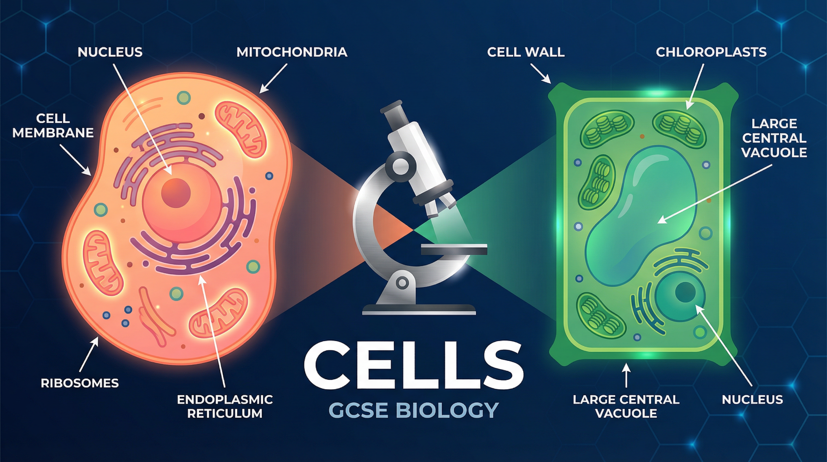
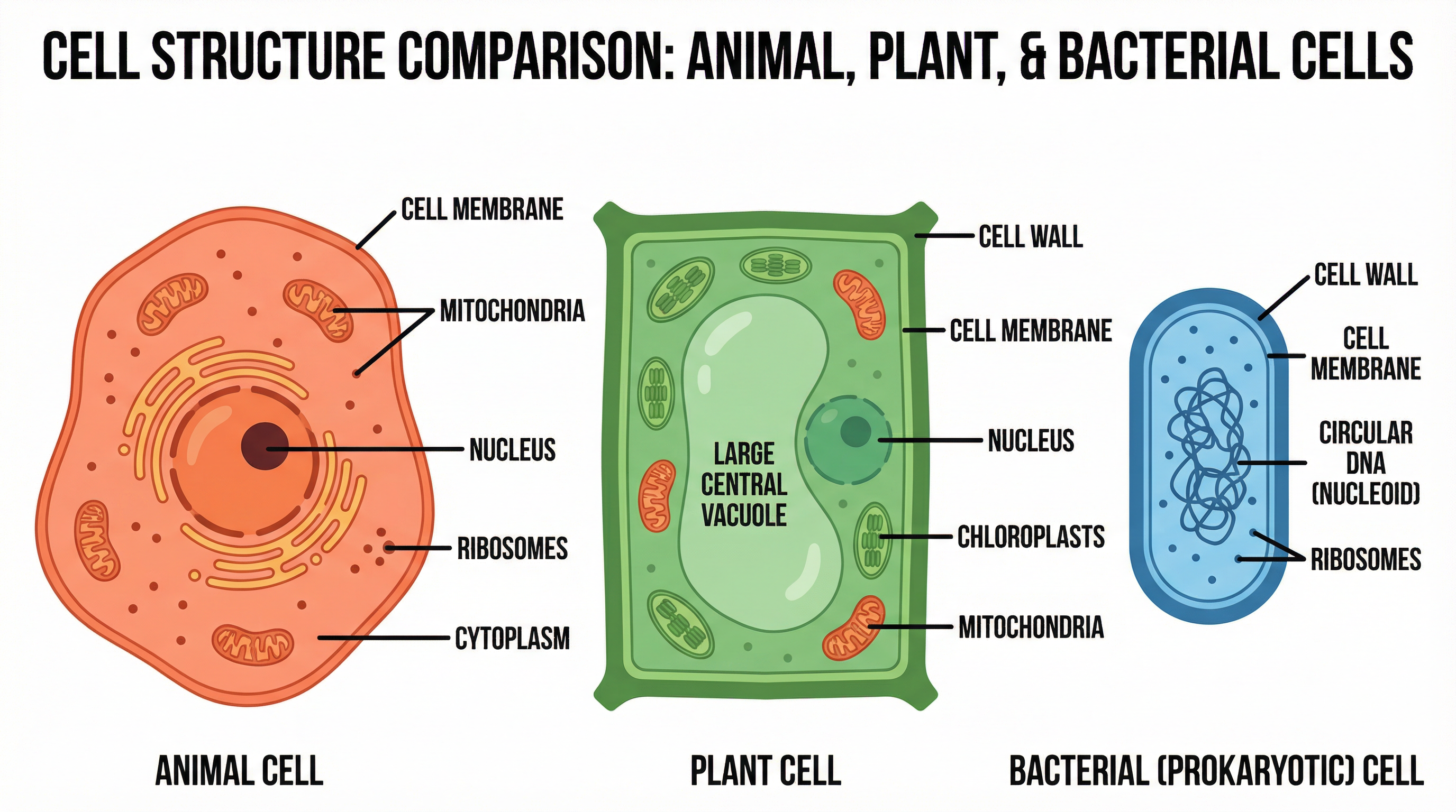
- Eukaryotic Cell
- A complex cell that contains a true, membrane-bound nucleus and other membrane-bound organelles.
- Prokaryotic Cell
- A smaller, simpler cell (like bacteria) that lacks a true nucleus; its genetic material floats freely in the cytoplasm.
- Differentiation
- The process by which a cell changes to become specialised for its job.
- Stem Cell
- An undifferentiated cell of an organism which is capable of giving rise to many more cells of the same type, and from which certain other cells can arise from differentiation.
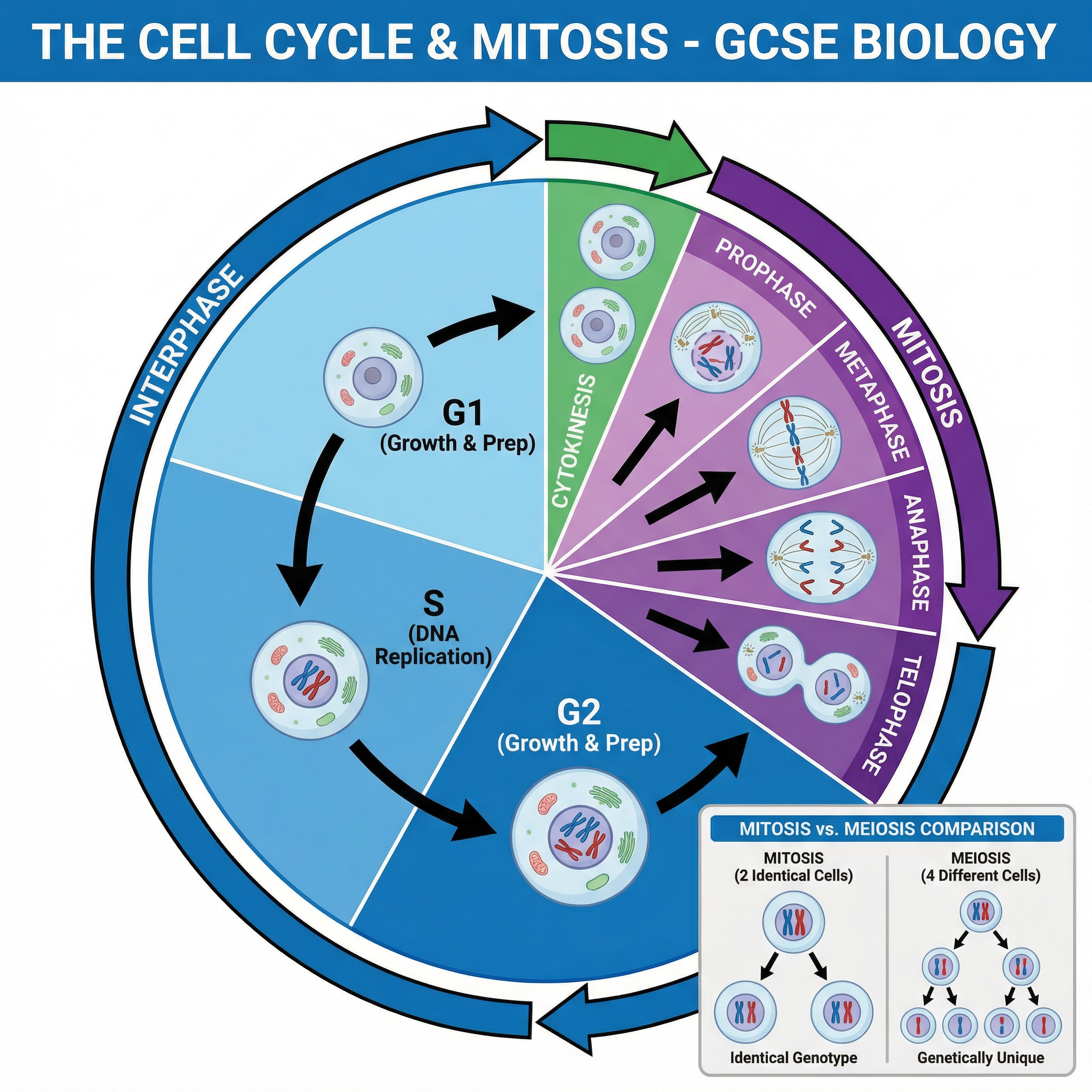
- Mitosis
- A type of cell division that results in two daughter cells each having the same number and kind of chromosomes as the parent nucleus, typical of ordinary tissue growth.
- Resolution
- The ability to distinguish between two separate points. The higher the resolution, the sharper the image.
Worked Examples
Worked Example
Question: A student measures the image of a cell in a textbook. The image is 45 mm wide. The magnification of the image is x1500. Calculate the actual width of the cell in micrometres (µm). Show your working.
Solution: Step 1: Convert the image size from mm to µm to match standard biological units. 45 mm × 1000 = 45,000 µm Step 2: State the formula. Actual Size = Image Size ÷ Magnification Step 3: Substitute the values into the formula. Actual Size = 45,000 ÷ 1500 Final answer: 30 µm
Worked Example
Question: Compare the structure of a red blood cell with the structure of a plant cell. (6 marks)
Solution: Differences: - A red blood cell has no nucleus, whereas a plant cell has a nucleus. - A red blood cell does not have a cell wall, whereas a plant cell has a cellulose cell wall. - A red blood cell has a biconcave disc shape, whereas a plant cell has a regular, fixed shape. - A red blood cell contains haemoglobin, which a plant cell does not. - A plant cell contains chloroplasts and a permanent vacuole, which a red blood cell does not. Similarities: - Both cells have a cell membrane. - Both cells contain cytoplasm.
Worked Example
Question: Describe what happens during the different stages of the cell cycle. (4 marks)
Solution: Stage 1 (Interphase): The cell grows and increases the number of sub-cellular structures such as ribosomes and mitochondria. The DNA replicates to form two copies of each chromosome. Stage 2 (Mitosis): One set of chromosomes is pulled to each end of the cell and the nucleus divides. Stage 3 (Cytokinesis): The cytoplasm and cell membrane divide to form two identical daughter cells.
Practice Questions
Question: Name two sub-cellular structures found in plant cells but not in animal cells.
Answer:
Question: Explain how a sperm cell is adapted for its function.
Answer:
Question: A student observed a plant cell using a light microscope. The actual length of the cell was 0.05 mm. The student drew the cell. The drawn cell was 20 mm long. Calculate the magnification of the drawing.
Answer:
Question: Evaluate the use of embryonic stem cells compared to adult stem cells in medical research and treatments.
Answer:
Question: Explain the advantages of using an electron microscope instead of a light microscope to view cells.
Answer: