AQA · A-Level · Biology
Course version: 7402 · Version from Sept 2016
Required practical 2: Preparation of stained squashes of cells from plant…
Full specification wording
All cells arise from other cells: Required practical 2: Preparation of stained squashes of cells from plant root tips; set-up and use of an optical microscope to identify the stages of mitosis in these stained squashes and calculation of a mitotic index.
Cut the last few millimetres from a growing root tip, as the meristem contains dividing cells.
Checked against the AQA 7402 specification (opens in a new tab) on .
- Point 14 of 14
- 4 key terms
Study notes
From the checked teaching notes for this specification point.
The idea
Cut the last few millimetres from a growing root tip, as the meristem contains dividing cells. Warm in 1 mol dm⁻³ hydrochloric acid at 60°C to hydrolyse the middle lamella, separating cells. Rinse, place on a slide, add a DNA stain (e.g. toluidine blue), cover with a coverslip and press firmly without twisting to squash cells into a single layer. Set up an optical microscope: start on the lowest objective lens, focus using the coarse knob, then switch to high power. Identify mitotic stages by chromosome appearance (e.g. chromosomes at equator in metaphase). Mitotic index = (cells in mitosis / total cells) × 100.
What earns the marks
- Use the root tip because it contains the meristem where dividing cells are located.
- Hydrolyse in hot hydrochloric acid to separate the cells.
- Squash to a single layer so that light can pass through the specimen.
- Stain the sample so that chromosomes become visible for identifying stages of mitosis.
- Calculate mitotic index as the number of cells in mitosis divided by the total number of cells counted.
Common mistakes and how to put them right
Counting only the dividing cells and forgetting that the denominator is every cell in the field, including those in interphase.
Explaining different mitotic indices as counting errors, rather than biological or sampling reasons such as different fields of view.
Twisting the coverslip during the squash, which tears cells instead of spreading them into one layer.
Exam tips
- Take a mean across several fields of view and state this; a single field is not representative.
- Show both counts used in the mitotic index calculation, not just the final figure.
Try the lesson
The start of the All cells arise from other cells: Required practical 2: Preparation of stained squashes of cells from plant root tips; set-up and use of an optical microscope to identify the stages of mitosis in these stained squashes and calculation of a mitotic index. lesson, made from checked teaching notes for this specification point.
Have a go first
Which part of a plant is used to prepare a stained squash for observing mitosis?
Listen
Cut the last few millimetres from a growing root tip, as the meristem contains dividing cells. Warm in 1 mol dm⁻³ hydrochloric acid at 60°C to hydrolyse the middle lamella, separating cells. Rinse, place on a slide, add a DNA stain (e.g. toluidine blue), cover with a coverslip and press firmly without twisting to squash cells into a single layer. Set up an optical microscope: start on the lowest objective lens, focus using the coarse knob, then switch to high power. Identify mitotic stages by chromosome appearance (e.g. chromosomes at equator in metaphase). Mitotic index = (cells in mitosis / total cells) × 100. A strong answer earns marks for these points. Use the root tip because it contains the meristem where dividing cells are located. Hydrolyse in hot hydrochloric acid to separate the cells. Squash to a single layer so that light can pass through the specimen. Stain the sample so that chromosomes become visible for identifying stages of mitosis. Exam tip: Take a mean across several fields of view and state this; a single field is not representative.
See it
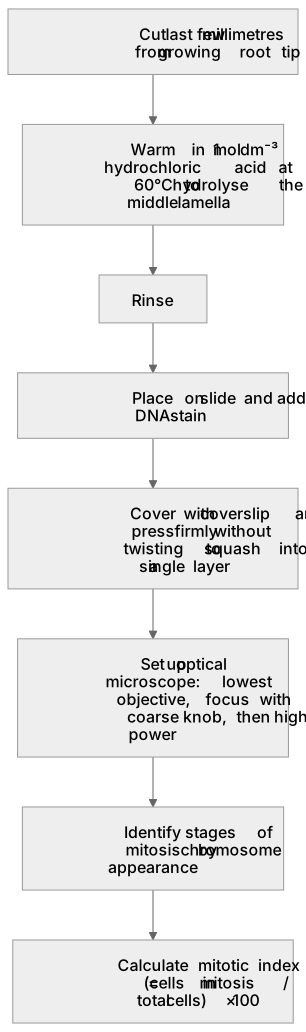
Put it in order
Put these steps in the order they happen.
- 1. Cover with coverslip and press firmly without twisting to squash into a single layer
- 2. Calculate mitotic index = (cells in mitosis / total cells) × 100
- 3. Set up optical microscope: lowest objective, focus with coarse knob, then high power
- 4. Cut last few millimetres from growing root tip
- 5. Warm in 1 mol dm⁻³ hydrochloric acid at 60°C to hydrolyse the middle lamella
- 6. Identify stages of mitosis by chromosome appearance
- 7. Rinse
- 8. Place on slide and add DNA stain
Key words
Say what each word means before you turn the card.
root tip meristem
Try answering aloud or in your head before turning the card.
acid hydrolysis and squashing
Try answering aloud or in your head before turning the card.
DNA stain
Try answering aloud or in your head before turning the card.
mitotic index
Try answering aloud or in your head before turning the card.
See every definition
- root tip meristem
- The part of the root containing dividing cells, which is why the last few millimetres of a growing root tip are cut.
- acid hydrolysis and squashing
- Warming in 1 mol dm⁻³ hydrochloric acid at 60°C hydrolyses the middle lamella to separate cells, and pressing firmly without twisting squashes cells into a single layer.
- DNA stain
- A stain such as toluidine blue added to the sample so that chromosomes become visible for identifying stages of mitosis.
- mitotic index
- Calculated as the number of cells in mitosis divided by the total number of cells counted, multiplied by 100.
Label the diagram
Some boxes have lost their names. Pick the right name for each number.
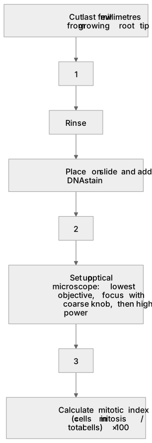
1Box 1
2Box 2
3Box 3
There is more to this lesson
The full lesson goes on to check your understanding and help you earn the marks. Create a free account to carry on, with your progress saved.
A week of Premium from your first lesson · Then the free plan continues · No card, no charge
Test your understanding of All cells arise from other cells: Required practical 2: Preparation of stained squashes of cells from plant root tips; set-up and use of an optical microscope to identify the stages of mitosis in these stained squashes and calculation of a mitotic index.
Active recall beats re-reading. Take an instant quiz now to test what you’ve learned and lock it into long-term memory.
A week of Premium from your first lesson · Then the free plan continues · No card, no charge