AQA · A-Level · Biology
Course version: 7402 · Version from Sept 2016
Cell recognition and the immune system: Antibody structure.
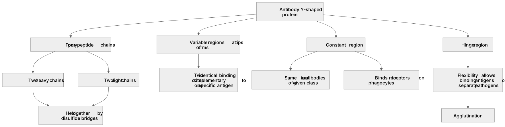
An antibody (such as IgG) is a Y-shaped protein with a quaternary structure comprising four polypeptide chains: two long heavy chains and two short light chains, held together by disulfide bridges.
Checked against the AQA 7402 specification (opens in a new tab) on .
- Point 8 of 18
- 3 key terms
Study notes
From the checked teaching notes for this specification point.
The idea
An antibody (such as IgG) is a Y-shaped protein with a quaternary structure comprising four polypeptide chains: two long heavy chains and two short light chains, held together by disulfide bridges. The tip of each arm is a variable region whose amino acid sequence and tertiary structure differ between antibodies. These form two identical binding sites complementary to one specific antigen. The rest of the molecule is the constant region, which is the same in all antibodies of a given class. This region binds to receptors on phagocytes, marking the antigen-antibody complex for engulfment. A flexible hinge region allows the arms to move apart and bind antigens on separate pathogens, causing agglutination.
What earns the marks
- The antibody has four polypeptide chains, two heavy and two light, giving it a quaternary structure.
- Disulfide bridges hold the polypeptide chains together.
- Two variable regions form two binding sites with a tertiary structure complementary to one specific antigen.
- The constant region is the same in all antibodies of a specific class and binds to receptors on phagocytes.
- The hinge region provides flexibility so that two separate antigens or pathogens can be bound simultaneously.
Common mistakes and how to put them right
Labelling the binding site as an active site.
Put it right: Antibodies are not enzymes.
Claiming the antibody changes shape to fit the antigen.
Put it right: Antibodies do not show induced fit.
Saying the variable region is the same in all antibodies.
Put it right: It is the constant region that is conserved within a class.
Drawing a single binding site, which makes explaining agglutination impossible.
Exam tips
- Label a sketch with variable region, binding site, constant region, hinge region, and disulfide bridge to clearly demonstrate structural knowledge.
- Pair every structural feature with its function, such as linking the presence of two binding sites to the ability to cause agglutination.
Try the lesson
The start of the Cell recognition and the immune system: Antibody structure. lesson, made from checked teaching notes for this specification point.
Have a go first
Which part of an antibody molecule binds to an antigen?
Listen
An antibody (such as IgG) is a Y-shaped protein with a quaternary structure comprising four polypeptide chains: two long heavy chains and two short light chains, held together by disulfide bridges. The tip of each arm is a variable region whose amino acid sequence and tertiary structure differ between antibodies. These form two identical binding sites complementary to one specific antigen. The rest of the molecule is the constant region, which is the same in all antibodies of a given class. This region binds to receptors on phagocytes, marking the antigen-antibody complex for engulfment. A flexible hinge region allows the arms to move apart and bind antigens on separate pathogens, causing agglutination. A strong answer earns marks for these points. The antibody has four polypeptide chains, two heavy and two light, giving it a quaternary structure. Disulfide bridges hold the polypeptide chains together. Two variable regions form two binding sites with a tertiary structure complementary to one specific antigen. The constant region is the same in all antibodies of a specific class and binds to receptors on phagocytes. Exam tip: Label a sketch with variable region, binding site, constant region, hinge region, and disulfide bridge to clearly demonstrate structural knowledge.
See it
Key words
Say what each word means before you turn the card.
variable region
Try answering aloud or in your head before turning the card.
constant region
Try answering aloud or in your head before turning the card.
disulfide bridges
Try answering aloud or in your head before turning the card.
See every definition
- variable region
- The tip of each arm, whose amino acid sequence and tertiary structure differ between antibodies and which forms a binding site complementary to one specific antigen.
- constant region
- The rest of the molecule, the same in all antibodies of a given class, which binds to receptors on phagocytes, marking the antigen-antibody complex for engulfment.
- disulfide bridges
- Bonds that hold the polypeptide chains of the antibody together.
Label the diagram
Some boxes have lost their names. Pick the right name for each number.
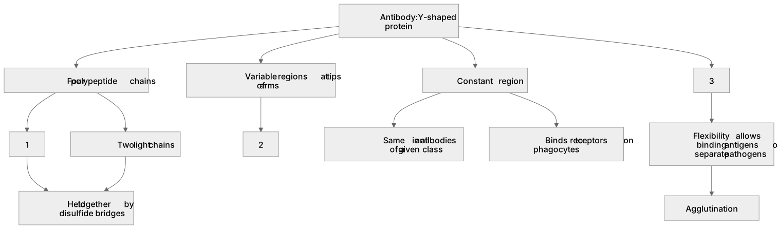
1Box 1
2Box 2
3Box 3
There is more to this lesson
The full lesson goes on to check your understanding and help you earn the marks. Create a free account to carry on, with your progress saved.
A week of Premium from your first lesson · Then the free plan continues · No card, no charge
Test your understanding of Cell recognition and the immune system: Antibody structure.
Active recall beats re-reading. Take an instant quiz now to test what you’ve learned and lock it into long-term memory.
A week of Premium from your first lesson · Then the free plan continues · No card, no charge