AQA · A-Level · Biology
Course version: 7402 · Version from Sept 2016
Measuring the size of an object viewed with an optical microscope.
Full specification wording
Methods of studying cells: Measuring the size of an object viewed with an optical microscope.
Size is measured with an eyepiece graticule, a scale etched into the eyepiece whose divisions have no fixed value until they are calibrated.
Checked against the AQA 7402 specification (opens in a new tab) on .
- Point 2 of 4
- 4 key terms
Study notes
From the checked teaching notes for this specification point.
The idea
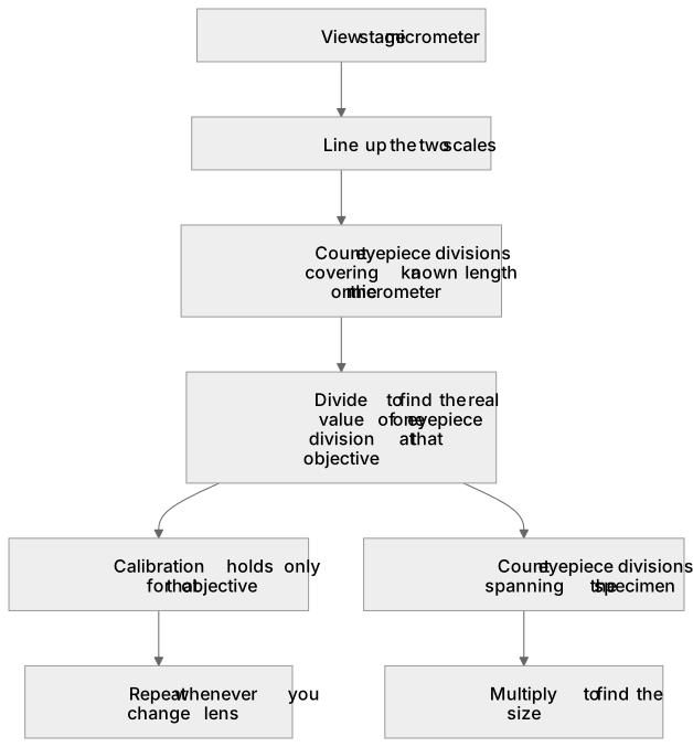
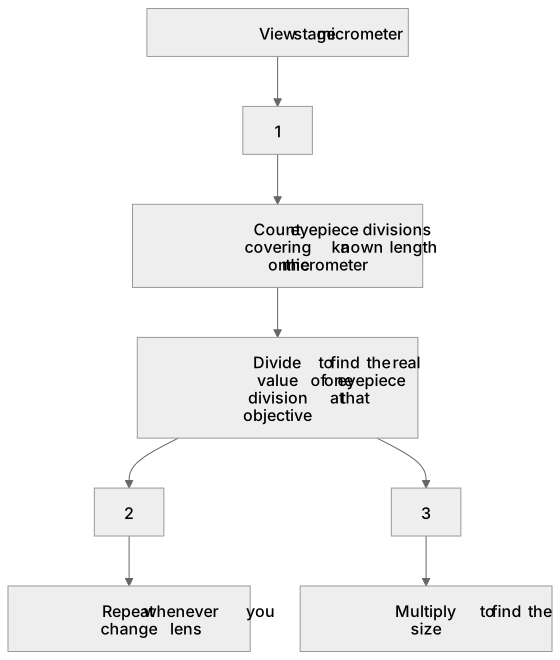
Size is measured with an eyepiece graticule, a scale etched into the eyepiece whose divisions have no fixed value until they are calibrated. You calibrate by viewing a stage micrometer, a slide carrying an accurate scale, usually one millimetre divided into one hundred divisions of ten micrometres. Line the two scales up, count how many eyepiece divisions cover a known length on the micrometer, and divide to find the real value of one eyepiece division at that objective. The calibration holds only for that objective, so it must be repeated whenever you change lens. Then count the eyepiece divisions spanning the specimen and multiply.
What earns the marks
- Measuring the image accurately and converting it to the same unit as the real object, for example 46 mm recorded as 46 000 micrometres.
- Correct use of magnification equals size of image divided by size of real object, shown as the division even if the arithmetic is wrong.
- A final magnification consistent with the measurement, quoted without units, for example about 20 000 times.
- Calibrating an eyepiece graticule against a stage micrometer, and recalibrating after changing the objective lens.
- Rearranging the formula correctly when the real size rather than the magnification is the unknown.
Common mistakes and how to put them right
Dividing the real size by the image size, which inverts the magnification.
Leaving the image in millimetres and the object in micrometres, so the answer is out by a factor of one thousand.
Attaching a unit to a magnification, which is a ratio and has none.
Treating eyepiece graticule divisions as fixed micrometres without calibrating against a stage micrometer.
Measuring the image carelessly.
Put it right: The mark scheme allows only a narrow band of ruler measurements, so a millimetre either way can cost the answer mark.
Exam tips
- Always show the conversion and the division; AQA awards a mark for correct use of the equation or the correct image size in micrometres even when the final answer is wrong.
- Convert everything to micrometres before dividing, then check the conversion rather than the size of the answer: a printed image has been enlarged far beyond what the microscope itself did, so an optical-microscope image of a bacterium can legitimately work out at about 20 000 times.
- Measure to the ends the question specifies, for instance excluding a flagellum if the stem says so.
Try the lesson
The start of the Methods of studying cells: Measuring the size of an object viewed with an optical microscope. lesson, made from checked teaching notes for this specification point.
Have a go first
A specimen is viewed at a total magnification of ×400. The image of a cell measures 20 mm. What is the actual length of the cell?
Listen
Size is measured with an eyepiece graticule, a scale etched into the eyepiece whose divisions have no fixed value until they are calibrated. You calibrate by viewing a stage micrometer, a slide carrying an accurate scale, usually one millimetre divided into one hundred divisions of ten micrometres. Line the two scales up, count how many eyepiece divisions cover a known length on the micrometer, and divide to find the real value of one eyepiece division at that objective. The calibration holds only for that objective, so it must be repeated whenever you change lens. Then count the eyepiece divisions spanning the specimen and multiply. A strong answer earns marks for these points. measuring the image accurately and converting it to the same unit as the real object, for example 46 mm recorded as 46 000 micrometres. correct use of magnification equals size of image divided by size of real object, shown as the division even if the arithmetic is wrong. a final magnification consistent with the measurement, quoted without units, for example about 20 000 times. calibrating an eyepiece graticule against a stage micrometer, and recalibrating after changing the objective lens. Exam tip: Always show the conversion and the division; AQA awards a mark for correct use of the equation or the correct image size in micrometres even when the final answer is wrong.
See it
Key words
Say what each word means before you turn the card.
eyepiece graticule
Try answering aloud or in your head before turning the card.
stage micrometer
Try answering aloud or in your head before turning the card.
magnification
Try answering aloud or in your head before turning the card.
calibration
Try answering aloud or in your head before turning the card.
See every definition
- eyepiece graticule
- A scale etched into the eyepiece whose divisions have no fixed value until they are calibrated.
- stage micrometer
- A slide carrying an accurate scale, usually one millimetre divided into one hundred divisions of ten micrometres.
- magnification
- Size of image divided by size of real object, a ratio that has no units.
- calibration
- Lining the two scales up, counting how many eyepiece divisions cover a known length on the micrometer, and dividing to find the real value of one eyepiece division at that objective.
Label the diagram
Some boxes have lost their names. Pick the right name for each number.
1Box 1
2Box 2
3Box 3
There is more to this lesson
The full lesson goes on to check your understanding and help you earn the marks. Create a free account to carry on, with your progress saved.
A week of Premium from your first lesson · Then the free plan continues · No card, no charge
Test your understanding of Methods of studying cells: Measuring the size of an object viewed with an optical microscope.
Active recall beats re-reading. Take an instant quiz now to test what you’ve learned and lock it into long-term memory.
A week of Premium from your first lesson · Then the free plan continues · No card, no charge