AQA · A-Level · Biology
Course version: 7402 · Version from Sept 2016
The use of marker genes to detect genetically modified (GM) cells or…
Full specification wording
Recombinant DNA technology (A-level only): The use of marker genes to detect genetically modified (GM) cells or organisms.
Only a small fraction of host cells take up a recombinant plasmid, so marker genes are used to identify them.
Checked against the AQA 7402 specification (opens in a new tab) on .
- Point 10 of 10
- 3 key terms
Study notes
From the checked teaching notes for this specification point.
The idea
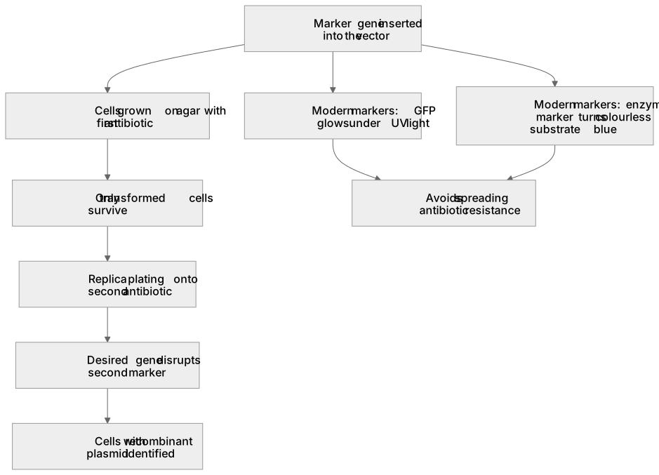
Only a small fraction of host cells take up a recombinant plasmid, so marker genes are used to identify them. A marker gene is inserted into the vector. However, plasmids can reseal without the gene of interest. Traditional methods use two antibiotic resistance genes. Cells are grown on agar with the first antibiotic; only transformed cells survive. Replica plating onto the second antibiotic identifies cells with the recombinant plasmid, as the desired gene disrupts the second marker. Modern markers avoid spreading antibiotic resistance: green fluorescent protein (GFP) makes recombinant cells glow under UV light, and enzyme markers like β-galactosidase turn a colourless substrate blue when the gene is intact.
What earns the marks
- The purpose of the marker gene: to show that the plasmid has been taken up, or to identify transformed cells.
- Explaining that the marker gene is inserted into the vector, allowing detection of cells that have taken up the plasmid.
- Naming a marker and its detection method, such as fluorescence under ultraviolet light or a colour change with an enzyme marker.
- Explaining how insertional inactivation (e.g., disrupting a second marker gene) distinguishes recombinant plasmids from empty plasmids.
Common mistakes and how to put them right
Saying the marker gene 'marks the gene', without stating that it identifies the cells that have taken up the plasmid.
Confusing cells with empty plasmids and recombinant plasmids.
Put it right: A single marker only shows plasmid uptake, while insertional inactivation of a second marker confirms the desired gene is present.
Naming the enzyme marker as lactase instead of β-galactosidase, or forgetting that fluorescent markers require ultraviolet light for detection.
Exam tips
- When asked how transformed cells are identified, state both the marker used and the specific method of detection (e.g., UV light for GFP).
- If asked for a modern alternative to antibiotic resistance, suggest fluorescent or enzyme markers and state that they prevent the spread of antibiotic resistance to pathogenic bacteria.
Try the lesson
The start of the Recombinant DNA technology (A-level only): The use of marker genes to detect genetically modified (GM) cells or organisms. lesson, made from checked teaching notes for this specification point.
Have a go first
What is the role of a marker gene in recombinant DNA technology?
Listen
Only a small fraction of host cells take up a recombinant plasmid, so marker genes are used to identify them. A marker gene is inserted into the vector. However, plasmids can reseal without the gene of interest. Traditional methods use two antibiotic resistance genes. Cells are grown on agar with the first antibiotic; only transformed cells survive. Replica plating onto the second antibiotic identifies cells with the recombinant plasmid, as the desired gene disrupts the second marker. Modern markers avoid spreading antibiotic resistance: green fluorescent protein (GFP) makes recombinant cells glow under UV light, and enzyme markers like β-galactosidase turn a colourless substrate blue when the gene is intact. A strong answer earns marks for these points. the purpose of the marker gene: to show that the plasmid has been taken up, or to identify transformed cells. explaining that the marker gene is inserted into the vector, allowing detection of cells that have taken up the plasmid. naming a marker and its detection method, such as fluorescence under ultraviolet light or a colour change with an enzyme marker. explaining how insertional inactivation (e.g., disrupting a second marker gene) distinguishes recombinant plasmids from empty plasmids. Exam tip: When asked how transformed cells are identified, state both the marker used and the specific method of detection (e.g., UV light for GFP).
See it
Key words
Say what each word means before you turn the card.
marker genes
Try answering aloud or in your head before turning the card.
identifying transformed cells
Try answering aloud or in your head before turning the card.
insertional inactivation
Try answering aloud or in your head before turning the card.
See every definition
- marker genes
- Genes inserted into the vector and used to detect genetically modified cells or organisms.
- identifying transformed cells
- Showing that the plasmid has been taken up, since only a small fraction of host cells take up a recombinant plasmid.
- insertional inactivation
- Disrupting a second marker gene, which distinguishes recombinant plasmids from empty plasmids because the desired gene disrupts the second marker.
Label the diagram
Some boxes have lost their names. Pick the right name for each number.
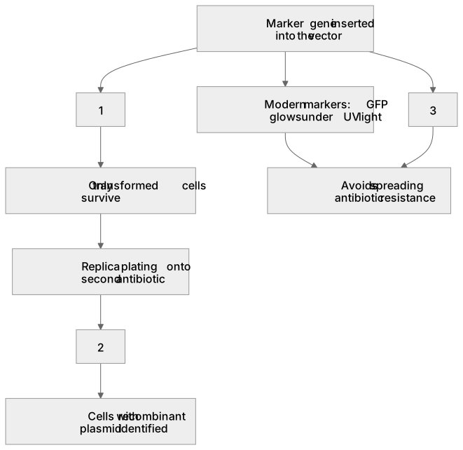
1Box 1
2Box 2
3Box 3
There is more to this lesson
The full lesson goes on to check your understanding and help you earn the marks. Create a free account to carry on, with your progress saved.
A week of Premium from your first lesson · Then the free plan continues · No card, no charge
Test your understanding of Recombinant DNA technology (A-level only): The use of marker genes to detect genetically modified (GM) cells or organisms.
Active recall beats re-reading. Take an instant quiz now to test what you’ve learned and lock it into long-term memory.
A week of Premium from your first lesson · Then the free plan continues · No card, no charge