AQA · A-Level · Biology
Course version: 7402 · Version from Sept 2016
Gross and microscopic structure of skeletal muscle. The ultrastructure of…
Full specification wording
Skeletal muscles are stimulated to contract by nerves and act as: Gross and microscopic structure of skeletal muscle. The ultrastructure of a myofibril.
A skeletal muscle is made of bundles of muscle fibres.
Checked against the AQA 7402 specification (opens in a new tab) on .
- Point 2 of 6
- 4 key terms
Study notes
From the checked teaching notes for this specification point.
The idea
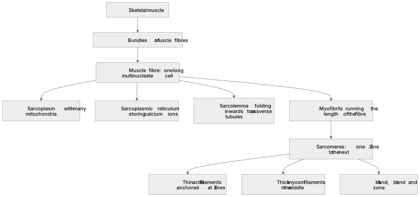
A skeletal muscle is made of bundles of muscle fibres. Each fibre is one very long cell formed when many cells fused, so it is multinucleate, with a shared cytoplasm called sarcoplasm containing many mitochondria and an extensive sarcoplasmic reticulum that stores calcium ions. Its plasma membrane, the sarcolemma, folds inwards at intervals as transverse tubules, which carry depolarisation deep into the fibre. Running the length of the fibre are myofibrils, each a chain of repeating units called sarcomeres. A sarcomere runs from one Z line to the next. The thin filaments are actin, anchored at the Z lines; the thick filaments are myosin, lying in the middle. The dark A band is the full length of the myosin, the light I band contains actin only, and the H zone is the part of the A band with myosin only. This banding is why skeletal muscle looks striated.
What earns the marks
- Describing a muscle fibre as multinucleate, containing sarcoplasm, many mitochondria and a sarcolemma.
- The sarcoplasmic reticulum storing calcium ions.
- Identifying a sarcomere as the region between two Z lines.
- Actin as the thin filament and myosin as the thick filament.
- Correctly identifying the A band, I band and H zone on a diagram or electron micrograph.
Common mistakes and how to put them right
Calling a muscle fibre a muscle, or a myofibril a fibre, when the hierarchy is muscle, fibre, myofibril, sarcomere, filament.
Placing actin in the middle of the sarcomere and myosin at the Z lines.
Assuming the A band contains actin because both begin with the same letter, when the A band is the dark band containing myosin.
Forgetting that the fibre is multinucleate, which follows from the fusion of many cells.
Labelling the sarcoplasmic reticulum without mentioning that it stores calcium ions.
Exam tips
- Learn the bands by what they contain, and the changes during contraction then follow automatically.
- On an electron micrograph find the Z lines first, then measure a sarcomere between them.
- Many marks depend on the calcium-storing role of the sarcoplasmic reticulum, so never name it without that function.
Try the lesson
The start of the Skeletal muscles are stimulated to contract by nerves and act as: Gross and microscopic structure of skeletal muscle. The ultrastructure of a myofibril. lesson, made from checked teaching notes for this specification point.
Have a go first
Which structure is the functional unit of a myofibril that shortens during muscle contraction?
Listen
A skeletal muscle is made of bundles of muscle fibres. Each fibre is one very long cell formed when many cells fused, so it is multinucleate, with a shared cytoplasm called sarcoplasm containing many mitochondria and an extensive sarcoplasmic reticulum that stores calcium ions. Its plasma membrane, the sarcolemma, folds inwards at intervals as transverse tubules, which carry depolarisation deep into the fibre. Running the length of the fibre are myofibrils, each a chain of repeating units called sarcomeres. A sarcomere runs from one Z line to the next. The thin filaments are actin, anchored at the Z lines; the thick filaments are myosin, lying in the middle. The dark A band is the full length of the myosin, the light I band contains actin only, and the H zone is the part of the A band with myosin only. This banding is why skeletal muscle looks striated. A strong answer earns marks for these points. describing a muscle fibre as multinucleate, containing sarcoplasm, many mitochondria and a sarcolemma. the sarcoplasmic reticulum storing calcium ions. identifying a sarcomere as the region between two Z lines. actin as the thin filament and myosin as the thick filament. Exam tip: Learn the bands by what they contain, and the changes during contraction then follow automatically.
See it
Key words
Say what each word means before you turn the card.
multinucleate fibre
Try answering aloud or in your head before turning the card.
sarcolemma and transverse tubules
Try answering aloud or in your head before turning the card.
sarcoplasmic reticulum
Try answering aloud or in your head before turning the card.
sarcomere between Z lines
Try answering aloud or in your head before turning the card.
See every definition
- multinucleate fibre
- One very long cell formed when many cells fused, with a shared cytoplasm called sarcoplasm containing many mitochondria and an extensive sarcoplasmic reticulum.
- sarcolemma and transverse tubules
- The plasma membrane, which folds inwards at intervals as transverse tubules that carry depolarisation deep into the fibre.
- sarcoplasmic reticulum
- An extensive structure in the sarcoplasm that stores calcium ions.
- sarcomere between Z lines
- A repeating unit of a myofibril, running from one Z line to the next, with thin actin filaments anchored at the Z lines and thick myosin filaments lying in the middle.
Label the diagram
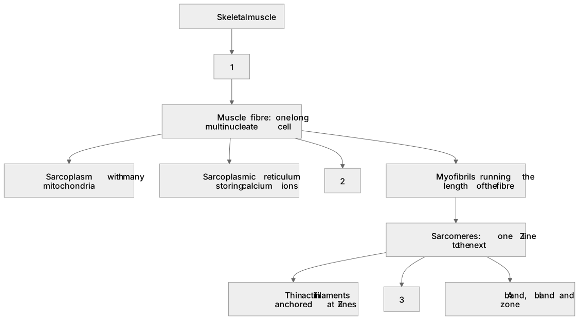
Some boxes have lost their names. Pick the right name for each number.
1Box 1
2Box 2
3Box 3
There is more to this lesson
The full lesson goes on to check your understanding and help you earn the marks. Create a free account to carry on, with your progress saved.
A week of Premium from your first lesson · Then the free plan continues · No card, no charge
Test your understanding of Skeletal muscles are stimulated to contract by nerves and act as: Gross and microscopic structure of skeletal muscle. The ultrastructure of a myofibril.
Active recall beats re-reading. Take an instant quiz now to test what you’ve learned and lock it into long-term memory.
A week of Premium from your first lesson · Then the free plan continues · No card, no charge