AQA · A-Level · Physics
Course version: 7408 · Version from Sept 2016
Basic principles of CT scanner: movement of X-ray tube narrow,…
Full specification wording
CT scanner (A-level only): Basic principles of CT scanner: movement of X-ray tube narrow, monochromatic X-ray beam array of detectors computer used to process the signals and produce a visual image.
A computed tomography (CT) scanner builds a cross-sectional image from many X-ray attenuation measurements.
Checked against the AQA 7408 specification (opens in a new tab) on .
- Point 1 of 2
- 3 key terms
Study notes
From the checked teaching notes for this specification point.
The idea
A computed tomography (CT) scanner builds a cross-sectional image from many X-ray attenuation measurements. A narrow, monochromatic X-ray beam is produced by an X-ray tube and collimated so that it passes through a thin slice of the patient. The tube and the detector array rotate around the patient, and the beam is also translated across the slice, so the same slice is viewed from many directions. Detectors measure the transmitted intensity after the beam has passed through the body; comparing this with the incident intensity gives the attenuation along each path. A computer processes the large set of path attenuation values, using reconstruction mathematics, to calculate the attenuation at each point in the slice and display it as a visual image. Dense tissue such as bone attenuates more than soft tissue, so contrast appears in the image.
What earns the marks
- Describes a narrow, monochromatic X-ray beam produced by an X-ray tube and collimated to pass through a thin slice of the patient.
- Explains that the X-ray tube and the detector array move around the patient, and that the beam is translated across the slice, so attenuation is measured along many different paths.
- States that detectors measure the transmitted intensity and that comparison with the incident intensity gives the attenuation along each beam path.
- Explains that a computer processes the many attenuation readings and reconstructs a cross-sectional visual image of the slice.
- Relates image contrast to differences in attenuation between tissues, for example bone attenuating more strongly than soft tissue.
- Recognises that using many directions avoids the ambiguity of a single projection, where overlapping tissues could give the same total attenuation.
Common mistakes and how to put them right
Saying the patient moves through the scanner while the tube and detectors stay fixed.
Put it right: In the basic description the tube and detector array rotate around the patient.
Describing a broad white beam rather than a narrow, monochromatic beam.
Put it right: The narrow beam defines the slice and monochromatic photons make attenuation depend on path length and material.
Claiming the computer takes a single photograph.
Put it right: The computer reconstructs an image from many separate attenuation measurements taken along different paths.
Confusing CT with a simple X-ray radiograph, where overlapping structures are superimposed rather than separated into a cross-sectional image.
Exam tips
- Use the sequence tube, collimation, transmission, detection, computer reconstruction, image when describing how a CT scan is produced.
- Name the components explicitly, such as X-ray tube, collimators, detector array and computer, to show precise knowledge.
- Explain why many directions are needed, linking the number of attenuation measurements to the ability to reconstruct a cross-section.
- Keep the description of the beam consistent: narrow and monochromatic, with attenuation measured along each path through the slice.
Try the lesson
The start of the CT scanner (A-level only): Basic principles of CT scanner: movement of X-ray tube narrow, monochromatic X-ray beam array of detectors computer used to process the signals and produce a visual image. lesson, made from checked teaching notes for this specification point.
Have a go first
In a CT scanner, what is the main role of the computer that receives signals from the detector array?
Listen
A computed tomography (CT) scanner builds a cross-sectional image from many X-ray attenuation measurements. A narrow, monochromatic X-ray beam is produced by an X-ray tube and collimated so that it passes through a thin slice of the patient. The tube and the detector array rotate around the patient, and the beam is also translated across the slice, so the same slice is viewed from many directions. Detectors measure the transmitted intensity after the beam has passed through the body; comparing this with the incident intensity gives the attenuation along each path. A computer processes the large set of path attenuation values, using reconstruction mathematics, to calculate the attenuation at each point in the slice and display it as a visual image. Dense tissue such as bone attenuates more than soft tissue, so contrast appears in the image. A strong answer earns marks for these points. Describes a narrow, monochromatic X-ray beam produced by an X-ray tube and collimated to pass through a thin slice of the patient. Explains that the X-ray tube and the detector array move around the patient, and that the beam is translated across the slice, so attenuation is measured along many different paths. States that detectors measure the transmitted intensity and that comparison with the incident intensity gives the attenuation along each beam path. Explains that a computer processes the many attenuation readings and reconstructs a cross-sectional visual image of the slice. Exam tip: Use the sequence tube, collimation, transmission, detection, computer reconstruction, image when describing how a CT scan is produced.
See it
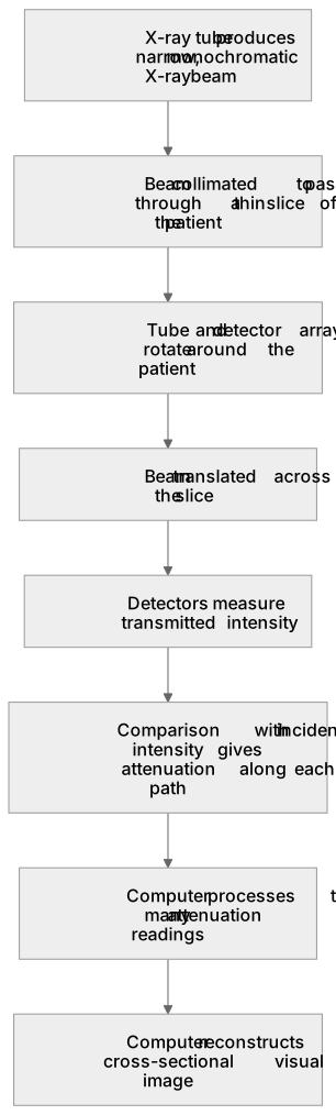
Put it in order
Put these steps in the order they happen.
- 1. Detectors measure transmitted intensity
- 2. Computer reconstructs a cross-sectional visual image
- 3. Comparison with incident intensity gives attenuation along each path
- 4. X-ray tube produces narrow, monochromatic X-ray beam
- 5. Beam collimated to pass through a thin slice of the patient
- 6. Computer processes the many attenuation readings
- 7. Tube and detector array rotate around the patient
- 8. Beam translated across the slice
Key words
Say what each word means before you turn the card.
Attenuation measurements along many paths
Try answering aloud or in your head before turning the card.
Computer reconstruction of cross-sectional images
Try answering aloud or in your head before turning the card.
Contrast from differences in tissue attenuation
Try answering aloud or in your head before turning the card.
See every definition
- Attenuation measurements along many paths
- Detectors measure the transmitted intensity after the beam has passed through the body, and comparing this with the incident intensity gives the attenuation along each path.
- Computer reconstruction of cross-sectional images
- A computer processes the large set of path attenuation values, using reconstruction mathematics, to calculate the attenuation at each point in the slice and display it as a visual image.
- Contrast from differences in tissue attenuation
- Dense tissue such as bone attenuates more than soft tissue, so contrast appears in the image.
Label the diagram
Some boxes have lost their names. Pick the right name for each number.
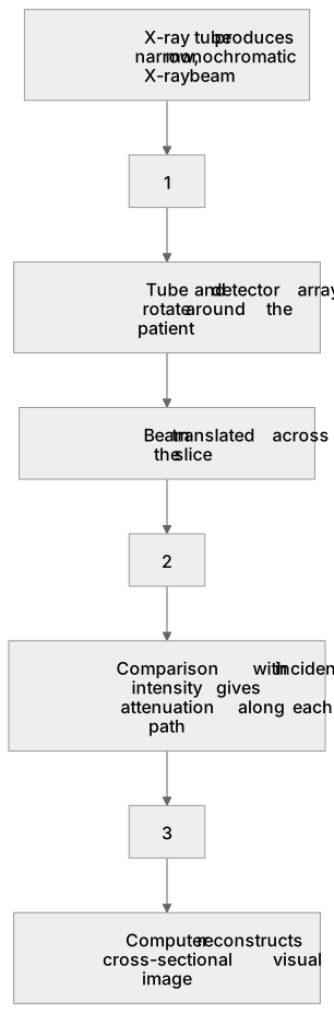
1Box 1
2Box 2
3Box 3
There is more to this lesson
The full lesson goes on to check your understanding and help you earn the marks. Create a free account to carry on, with your progress saved.
A week of Premium from your first lesson · Then the free plan continues · No card, no charge
Test your understanding of CT scanner (A-level only): Basic principles of CT scanner: movement of X-ray tube narrow, monochromatic X-ray beam array of detectors computer used to process the signals and produce a visual image.
Active recall beats re-reading. Take an instant quiz now to test what you’ve learned and lock it into long-term memory.
A week of Premium from your first lesson · Then the free plan continues · No card, no charge