AQA · A-Level · Physics
Course version: 7408 · Version from Sept 2016
Principle of operation of the transmission electron microscope (TEM).
Full specification wording
Electron microscopes (A-level only): Principle of operation of the transmission electron microscope (TEM).
A TEM uses a beam of accelerated electrons, produced by an electron gun and focused by magnetic lenses, to illuminate a very thin specimen.
Checked against the AQA 7408 specification (opens in a new tab) on .
- Point 2 of 3
- 4 key terms
Study notes
From the checked teaching notes for this specification point.
The idea
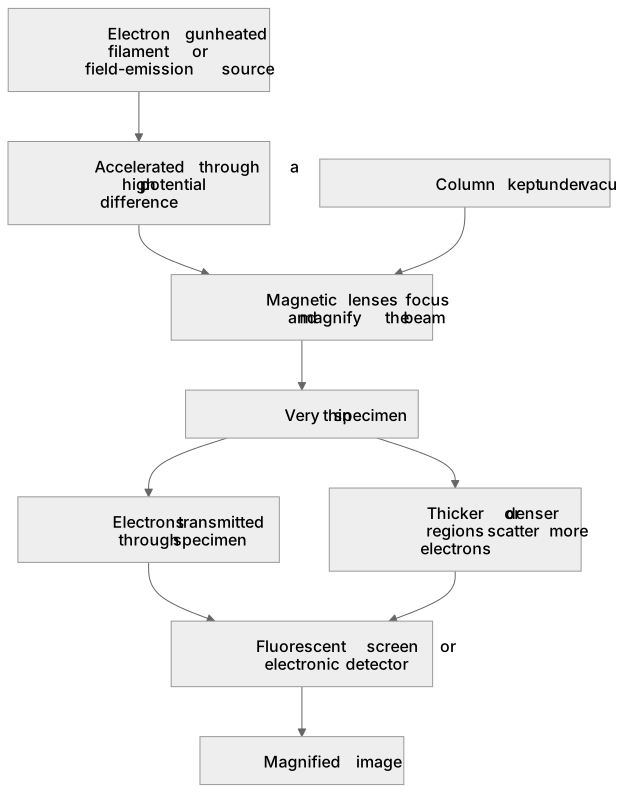
A TEM uses a beam of accelerated electrons, produced by an electron gun and focused by magnetic lenses, to illuminate a very thin specimen. Because electrons have a much shorter de Broglie wavelength than visible light, the resolution is far better than an optical microscope. Electrons pass through the specimen; regions that are thicker or denser scatter more electrons, so fewer reach the fluorescent screen or detector, forming a magnified image. The whole column is evacuated so electrons are not scattered by air. The specimen must be extremely thin so electrons can transmit through it. Contrast arises from differences in electron scattering, and the image can be recorded digitally or on film.
What earns the marks
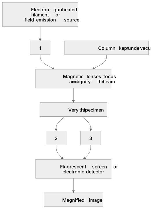
- Electrons are emitted from a heated filament or field-emission source and accelerated through a high potential difference, giving them high kinetic energy and a short de Broglie wavelength.
- Magnetic lenses, not glass lenses, focus and magnify the electron beam because glass would absorb or scatter electrons.
- The specimen must be very thin so that a sufficient number of electrons can pass through it rather than being absorbed or scattered.
- Electrons that pass through the specimen are detected on a fluorescent screen or by an electronic detector, producing a magnified image.
- Contrast is produced because thicker or denser regions scatter electrons more, so fewer electrons reach the detector from those areas.
- The column is kept under vacuum to prevent electrons from colliding with gas molecules, which would scatter the beam and blur the image.
Common mistakes and how to put them right
Thinking that glass lenses are used to focus electrons.
Put it right: Magnetic lenses are used because glass would absorb or scatter electrons.
Believing that the image is formed by electrons reflected from the surface.
Put it right: In a TEM electrons are transmitted through a very thin specimen.
Assuming that a TEM can examine any solid sample directly.
Put it right: Specimens must be extremely thin and prepared specially, and the column must be evacuated.
Exam tips
- Link the high resolution explicitly to the short de Broglie wavelength of accelerated electrons, not simply to their high speed.
- Describe the path of electrons in order: source, acceleration, magnetic lenses, thin specimen, detector.
- When comparing with an optical microscope, mention both the wavelength advantage and the need for a vacuum and thin specimens.
Try the lesson
The start of the Electron microscopes (A-level only): Principle of operation of the transmission electron microscope (TEM). lesson, made from checked teaching notes for this specification point.
Have a go first
In a transmission electron microscope, what happens to the electrons after they pass through the specimen?
Listen
A TEM uses a beam of accelerated electrons, produced by an electron gun and focused by magnetic lenses, to illuminate a very thin specimen. Because electrons have a much shorter de Broglie wavelength than visible light, the resolution is far better than an optical microscope. Electrons pass through the specimen; regions that are thicker or denser scatter more electrons, so fewer reach the fluorescent screen or detector, forming a magnified image. The whole column is evacuated so electrons are not scattered by air. The specimen must be extremely thin so electrons can transmit through it. Contrast arises from differences in electron scattering, and the image can be recorded digitally or on film. A strong answer earns marks for these points. Electrons are emitted from a heated filament or field-emission source and accelerated through a high potential difference, giving them high kinetic energy and a short de Broglie wavelength. Magnetic lenses, not glass lenses, focus and magnify the electron beam because glass would absorb or scatter electrons. The specimen must be very thin so that a sufficient number of electrons can pass through it rather than being absorbed or scattered. Electrons that pass through the specimen are detected on a fluorescent screen or by an electronic detector, producing a magnified image. A common slip: Thinking that glass lenses are used to focus electrons. Instead, magnetic lenses are used because glass would absorb or scatter electrons. Exam tip: Link the high resolution explicitly to the short de Broglie wavelength of accelerated electrons, not simply to their high speed.
See it
Key words
Say what each word means before you turn the card.
Wave-particle duality and the de Broglie wavelength
Try answering aloud or in your head before turning the card.
Electron optics and magnetic focusing
Try answering aloud or in your head before turning the card.
Resolution limits of microscopes
Try answering aloud or in your head before turning the card.
Specimen preparation and vacuum requirements
Try answering aloud or in your head before turning the card.
See every definition
- Wave-particle duality and the de Broglie wavelength
- Accelerated electrons have a much shorter de Broglie wavelength than visible light, which gives a far better resolution than an optical microscope.
- Electron optics and magnetic focusing
- Magnetic lenses focus and magnify the electron beam because glass would absorb or scatter electrons.
- Resolution limits of microscopes
- Because electrons have a much shorter de Broglie wavelength than visible light, the resolution is far better than an optical microscope.
- Specimen preparation and vacuum requirements
- The specimen must be extremely thin so electrons can transmit through it, and the whole column is evacuated so electrons are not scattered by air.
Label the diagram
Some boxes have lost their names. Pick the right name for each number.
1Box 1
2Box 2
3Box 3
There is more to this lesson
The full lesson goes on to check your understanding and help you earn the marks. Create a free account to carry on, with your progress saved.
A week of Premium from your first lesson · Then the free plan continues · No card, no charge
Test your understanding of Electron microscopes (A-level only): Principle of operation of the transmission electron microscope (TEM).
Active recall beats re-reading. Take an instant quiz now to test what you’ve learned and lock it into long-term memory.
A week of Premium from your first lesson · Then the free plan continues · No card, no charge