AQA · A-Level · Physics
Course version: 7408 · Version from Sept 2016
Photographic detection with intensifying screen and fluoroscopic image…
Full specification wording
Image detection and enhancement (A-level only): Photographic detection with intensifying screen and fluoroscopic image intensification; reasons for using these.
X-ray film blackens when X-ray photons interact with the emulsion, but direct exposure needs a large dose because film responds weakly to X-rays.
Checked against the AQA 7408 specification (opens in a new tab) on .
- Point 4 of 4
- 4 key terms
Study notes
From the checked teaching notes for this specification point.
The idea
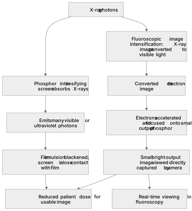
X-ray film blackens when X-ray photons interact with the emulsion, but direct exposure needs a large dose because film responds weakly to X-rays. An intensifying screen contains a phosphor that absorbs X-ray photons and emits many visible photons, which expose the film far more efficiently. The screen is placed in contact with the film, so one absorbed X-ray photon produces a local burst of light and a darker spot. Fluoroscopic image intensification instead converts the X-ray pattern into visible light, then into electrons, accelerates and focuses them, and produces a small bright output image that a camera or screen can display. Both methods reduce the patient dose needed for a usable image and allow real-time viewing in fluoroscopy.
What earns the marks
- Photographic detection uses film whose emulsion is blackened by radiation, but direct X-ray exposure is inefficient and requires a relatively large dose.
- An intensifying screen contains a phosphor that absorbs X-ray photons and emits visible or ultraviolet photons, which expose the film more effectively.
- The screen is used in close contact with the film so that light from each X-ray interaction spreads only a small distance, preserving detail while amplifying the signal.
- Fluoroscopic image intensification converts the X-ray image into a visible-light image, then into an electron image that is accelerated and focused onto a small output phosphor.
- The output image is much brighter than the original fluoroscopic screen image, so it can be viewed directly or captured by a camera, including in real time.
Common mistakes and how to put them right
Stating that the intensifying screen absorbs visible light and re-emits X-rays.
Put it right: It absorbs X-rays and emits visible or ultraviolet light.
Confusing image intensification with contrast enhancement.
Put it right: Intensification increases the brightness or signal of the image, whereas contrast media change the attenuation of the subject.
Claiming that intensification removes the need for any X-ray dose.
Put it right: It reduces the dose needed for a usable image but X-rays are still required to form the initial pattern.
Exam tips
- Describe the energy conversion sequence in each device in order, naming the input and output radiation at each stage.
- Explain the benefit in terms of signal per unit dose and, for fluoroscopy, the ability to view the image in real time.
- Distinguish clearly between intensifying screens used with film and image intensifiers used in fluoroscopy.
Try the lesson
The start of the Image detection and enhancement (A-level only): Photographic detection with intensifying screen and fluoroscopic image intensification; reasons for using these. lesson, made from checked teaching notes for this specification point.
Have a go first
Why is an intensifying screen placed next to the photographic film in a radiography cassette?
Listen
X-ray film blackens when X-ray photons interact with the emulsion, but direct exposure needs a large dose because film responds weakly to X-rays. An intensifying screen contains a phosphor that absorbs X-ray photons and emits many visible photons, which expose the film far more efficiently. The screen is placed in contact with the film, so one absorbed X-ray photon produces a local burst of light and a darker spot. Fluoroscopic image intensification instead converts the X-ray pattern into visible light, then into electrons, accelerates and focuses them, and produces a small bright output image that a camera or screen can display. Both methods reduce the patient dose needed for a usable image and allow real-time viewing in fluoroscopy. A strong answer earns marks for these points. Photographic detection uses film whose emulsion is blackened by radiation, but direct X-ray exposure is inefficient and requires a relatively large dose. An intensifying screen contains a phosphor that absorbs X-ray photons and emits visible or ultraviolet photons, which expose the film more effectively. The screen is used in close contact with the film so that light from each X-ray interaction spreads only a small distance, preserving detail while amplifying the signal. Fluoroscopic image intensification converts the X-ray image into a visible-light image, then into an electron image that is accelerated and focused onto a small output phosphor. A common slip: Stating that the intensifying screen absorbs visible light and re-emits X-rays. Instead, it absorbs X-rays and emits visible or ultraviolet light. Exam tip: Describe the energy conversion sequence in each device in order, naming the input and output radiation at each stage.
See it
Key words
Say what each word means before you turn the card.
intensifying screen
Try answering aloud or in your head before turning the card.
fluoroscopic image intensification
Try answering aloud or in your head before turning the card.
dose reduction
Try answering aloud or in your head before turning the card.
real-time viewing
Try answering aloud or in your head before turning the card.
See every definition
- intensifying screen
- Contains a phosphor that absorbs X-ray photons and emits many visible photons, which expose the film far more efficiently.
- fluoroscopic image intensification
- Converts the X-ray pattern into visible light, then into electrons, accelerates and focuses them, and produces a small bright output image.
- dose reduction
- Both methods increase the signal produced per unit patient dose, so they reduce the dose required for a diagnostic image.
- real-time viewing
- Fluoroscopic image intensification produces a bright output image that a camera or screen can display, allowing real-time viewing in fluoroscopy.
Label the diagram
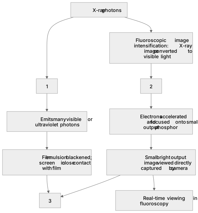
Some boxes have lost their names. Pick the right name for each number.
1Box 1
2Box 2
3Box 3
There is more to this lesson
The full lesson goes on to check your understanding and help you earn the marks. Create a free account to carry on, with your progress saved.
A week of Premium from your first lesson · Then the free plan continues · No card, no charge
Test your understanding of Image detection and enhancement (A-level only): Photographic detection with intensifying screen and fluoroscopic image intensification; reasons for using these.
Active recall beats re-reading. Take an instant quiz now to test what you’ve learned and lock it into long-term memory.
A week of Premium from your first lesson · Then the free plan continues · No card, no charge