AQA · A-Level · Physics
Course version: 7408 · Version from Sept 2016
Rotating-anode X-ray tube; methods of controlling the beam intensity, the…
Full specification wording
The physics of diagnostic X-rays(A-level only): Rotating-anode X-ray tube; methods of controlling the beam intensity, the photon energy, the image sharpness and contrast, and the patient dose.
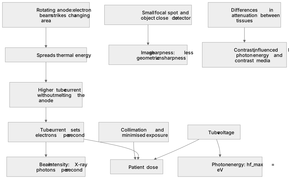
A rotating-anode X-ray tube spins a bevelled metal target so the electron beam strikes a changing area, spreading thermal energy and allowing a higher beam current without melting the anode.
Checked against the AQA 7408 specification (opens in a new tab) on .
- Point 2 of 2
- 4 key terms
Study notes
From the checked teaching notes for this specification point.
The idea
A rotating-anode X-ray tube spins a bevelled metal target so the electron beam strikes a changing area, spreading thermal energy and allowing a higher beam current without melting the anode. Beam intensity is controlled by the tube current, which sets the number of electrons per second and hence the number of X-ray photons. Photon energy is controlled by the tube voltage, since hf_max = eV. Image sharpness improves when the focal spot is small and the object is close to the detector, reducing geometric unsharpness. Contrast depends on differences in attenuation between tissues, which is influenced by photon energy and the use of contrast media. Patient dose rises with tube current, exposure time and tube voltage, so the beam is collimated and exposure is minimised.
What earns the marks
- The anode rotates so the electron beam strikes a moving area, preventing local overheating and allowing a higher tube current and continuous operation.
- Tube current controls beam intensity: a larger current means more electrons per second and therefore more X-ray photons per second.
- Tube voltage controls photon energy: increasing V increases hf_max = eV and shifts the spectrum to higher energies.
- Image sharpness is improved by a small focal spot and by reducing the object-to-detector distance, which reduces geometric blurring.
- Contrast depends on differential attenuation; lower-energy photons generally increase contrast between soft tissues but also increase patient dose.
- Patient dose is reduced by collimating the beam, minimising exposure time, using appropriate filtration and avoiding unnecessary repeat exposures.
Common mistakes and how to put them right
Saying tube current controls photon energy.
Put it right: Tube current controls intensity, while tube voltage controls photon energy.
Ignoring the effect of focal spot size on sharpness.
Put it right: A smaller focal spot reduces geometric unsharpness and improves sharpness.
Assuming higher photon energy always improves contrast.
Put it right: Higher energy can reduce contrast because attenuation differences between tissues become smaller.
Exam tips
- Link each control to a specific physical quantity: current to intensity, voltage to photon energy, collimation and time to dose.
- When discussing sharpness, refer to focal spot size and distances rather than vague statements about image quality.
- For contrast, explain differential attenuation and mention how photon energy or contrast media change it.
Try the lesson
The start of the The physics of diagnostic X-rays(A-level only): Rotating-anode X-ray tube; methods of controlling the beam intensity, the photon energy, the image sharpness and contrast, and the patient dose. lesson, made from checked teaching notes for this specification point.
Have a go first
Why is the anode in a diagnostic X-ray tube rotated during operation?
Listen
A rotating-anode X-ray tube spins a bevelled metal target so the electron beam strikes a changing area, spreading thermal energy and allowing a higher beam current without melting the anode. Beam intensity is controlled by the tube current, which sets the number of electrons per second and hence the number of X-ray photons. Photon energy is controlled by the tube voltage, since hf_max = eV. Image sharpness improves when the focal spot is small and the object is close to the detector, reducing geometric unsharpness. Contrast depends on differences in attenuation between tissues, which is influenced by photon energy and the use of contrast media. Patient dose rises with tube current, exposure time and tube voltage, so the beam is collimated and exposure is minimised. A strong answer earns marks for these points. The anode rotates so the electron beam strikes a moving area, preventing local overheating and allowing a higher tube current and continuous operation. Tube current controls beam intensity: a larger current means more electrons per second and therefore more X-ray photons per second. Tube voltage controls photon energy: increasing V increases hf_max = eV and shifts the spectrum to higher energies. Image sharpness is improved by a small focal spot and by reducing the object-to-detector distance, which reduces geometric blurring. A common slip: Saying tube current controls photon energy. Instead, tube current controls intensity, while tube voltage controls photon energy. Exam tip: Link each control to a specific physical quantity: current to intensity, voltage to photon energy, collimation and time to dose.
See it
Key words
Say what each word means before you turn the card.
Thermal management in X-ray tubes
Try answering aloud or in your head before turning the card.
Control of beam intensity and photon energy
Try answering aloud or in your head before turning the card.
Image quality: sharpness and contrast
Try answering aloud or in your head before turning the card.
Radiation protection and patient dose
Try answering aloud or in your head before turning the card.
See every definition
- Thermal management in X-ray tubes
- The anode rotates so the electron beam strikes a moving area, preventing local overheating and allowing a higher tube current and continuous operation.
- Control of beam intensity and photon energy
- Tube current controls beam intensity, while tube voltage controls photon energy, since increasing V increases hf_max = eV.
- Image quality: sharpness and contrast
- Sharpness is improved by a small focal spot and reduced object-to-detector distance, while contrast depends on differential attenuation.
- Radiation protection and patient dose
- Dose is reduced by collimating the beam, minimising exposure time, using appropriate filtration and avoiding unnecessary repeat exposures.
Label the diagram
Some boxes have lost their names. Pick the right name for each number.
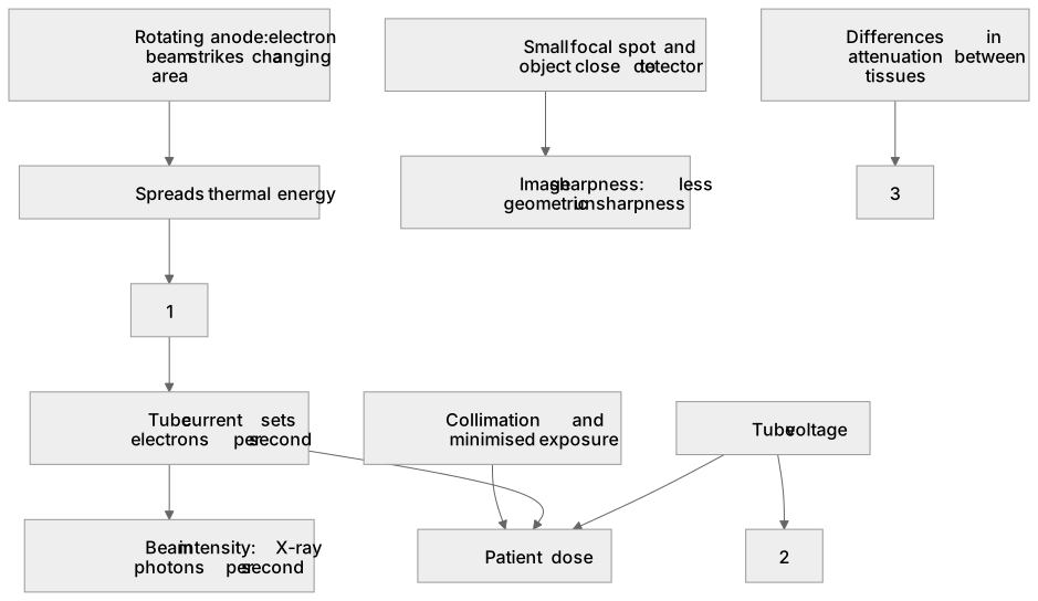
1Box 1
2Box 2
3Box 3
There is more to this lesson
The full lesson goes on to check your understanding and help you earn the marks. Create a free account to carry on, with your progress saved.
A week of Premium from your first lesson · Then the free plan continues · No card, no charge
Test your understanding of The physics of diagnostic X-rays(A-level only): Rotating-anode X-ray tube; methods of controlling the beam intensity, the photon energy, the image sharpness and contrast, and the patient dose.
Active recall beats re-reading. Take an instant quiz now to test what you’ve learned and lock it into long-term memory.
A week of Premium from your first lesson · Then the free plan continues · No card, no charge