AQA · A-Level · Biology
Course version: 7402 · Version from Sept 2016
The structure of arteries, arterioles and veins in relation to their…
Full specification wording
Mass transport in animals: The structure of arteries, arterioles and veins in relation to their function.
All three vessel types share an inner endothelium, a middle layer of smooth muscle and elastic tissue, and an outer collagen coat, but the proportions differ with function.
Checked against the AQA 7402 specification (opens in a new tab) on .
- Point 11 of 14
- 4 key terms
Study notes
From the checked teaching notes for this specification point.
The idea
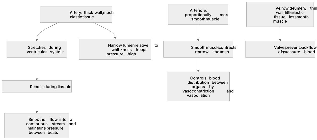
All three vessel types share an inner endothelium, a middle layer of smooth muscle and elastic tissue, and an outer collagen coat, but the proportions differ with function. Arteries carry blood at high pressure away from the heart, so they have thick walls with much elastic tissue: it stretches as blood is forced in during ventricular systole and recoils during diastole, smoothing flow into a continuous stream and maintaining pressure between beats. A narrow lumen relative to wall thickness keeps pressure high. Arterioles have proportionally more smooth muscle, which contracts to narrow the lumen, controlling blood distribution between organs by vasoconstriction and vasodilation. Veins carry blood at low pressure back to the heart, so they have a wide lumen, a thin wall with little elastic tissue and less smooth muscle, and valves that prevent backflow.
What earns the marks
- A thick artery wall containing elastic tissue enabling the vessel to withstand and maintain high pressure.
- Elastic recoil of the artery wall smoothing out the pulse and maintaining flow during diastole.
- Smooth muscle in arterioles contracting to narrow the lumen and redirect blood flow.
- A vein having a wide lumen and valves to prevent backflow of low pressure blood.
- A vein having a thin wall with little elastic tissue because the blood it carries is at low pressure.
Common mistakes and how to put them right
Writing thick muscular walls for arteries when the mark is for elastic tissue and pressure.
Saying veins have valves because blood moves slowly, without mentioning backflow or low pressure.
Confusing the lumen with the wall, so a vein is described as having a narrow lumen.
Claiming arteries contain valves, or that arterioles contain no muscle.
Listing a structure with no function attached, which scores nothing in an in relation to their function question.
Exam tips
- Write every point as structure, then because, then function, in one sentence.
- Use the word lumen, and say relative to the wall thickness when you compare vessels.
- Remember it is the arterioles, not the arteries, that control the distribution of blood between organs.
Try the lesson
The start of the Mass transport in animals: The structure of arteries, arterioles and veins in relation to their function. lesson, made from checked teaching notes for this specification point.
Have a go first
Which structural feature of an artery is directly responsible for withstanding the high pressure of blood leaving the heart?
Listen
All three vessel types share an inner endothelium, a middle layer of smooth muscle and elastic tissue, and an outer collagen coat, but the proportions differ with function. Arteries carry blood at high pressure away from the heart, so they have thick walls with much elastic tissue: it stretches as blood is forced in during ventricular systole and recoils during diastole, smoothing flow into a continuous stream and maintaining pressure between beats. A narrow lumen relative to wall thickness keeps pressure high. Arterioles have proportionally more smooth muscle, which contracts to narrow the lumen, controlling blood distribution between organs by vasoconstriction and vasodilation. Veins carry blood at low pressure back to the heart, so they have a wide lumen, a thin wall with little elastic tissue and less smooth muscle, and valves that prevent backflow. A strong answer earns marks for these points. a thick artery wall containing elastic tissue enabling the vessel to withstand and maintain high pressure. elastic recoil of the artery wall smoothing out the pulse and maintaining flow during diastole. smooth muscle in arterioles contracting to narrow the lumen and redirect blood flow. a vein having a wide lumen and valves to prevent backflow of low pressure blood. Exam tip: Write every point as structure, then because, then function, in one sentence.
See it
Key words
Say what each word means before you turn the card.
elastic recoil
Try answering aloud or in your head before turning the card.
high and low pressure vessels
Try answering aloud or in your head before turning the card.
lumen diameter
Try answering aloud or in your head before turning the card.
smooth muscle and vasoconstriction
Try answering aloud or in your head before turning the card.
See every definition
- elastic recoil
- The elastic tissue in an artery wall stretches as blood is forced in during ventricular systole and recoils during diastole, smoothing flow into a continuous stream and maintaining pressure between beats.
- high and low pressure vessels
- Arteries carry blood at high pressure away from the heart, while veins carry blood at low pressure back to the heart.
- lumen diameter
- A narrow lumen relative to wall thickness keeps pressure high, whereas a vein has a wide lumen.
- smooth muscle and vasoconstriction
- Arterioles have proportionally more smooth muscle, which contracts to narrow the lumen, controlling blood distribution between organs by vasoconstriction and vasodilation.
Label the diagram
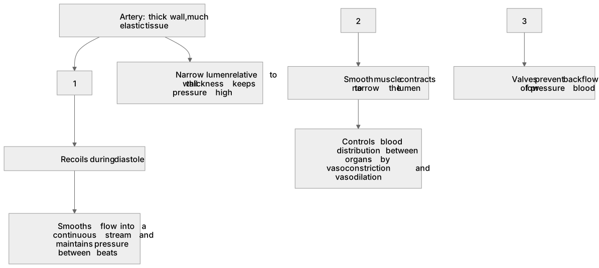
Some boxes have lost their names. Pick the right name for each number.
1Box 1
2Box 2
3Box 3
There is more to this lesson
The full lesson goes on to check your understanding and help you earn the marks. Create a free account to carry on, with your progress saved.
A week of Premium from your first lesson · Then the free plan continues · No card, no charge
Test your understanding of Mass transport in animals: The structure of arteries, arterioles and veins in relation to their function.
Active recall beats re-reading. Take an instant quiz now to test what you’ve learned and lock it into long-term memory.
A week of Premium from your first lesson · Then the free plan continues · No card, no charge