AQA · A-Level · Biology
Course version: 7402 · Version from Sept 2016
The roles of calcium ions and tropomyosin in the cycle of actinomyosin…
Full specification wording
Skeletal muscles are stimulated to contract by nerves and act as: The roles of calcium ions and tropomyosin in the cycle of actinomyosin bridge formation.
In relaxed muscle, tropomyosin covers myosin binding sites on actin filaments, preventing actinomyosin bridge formation.
Checked against the AQA 7402 specification (opens in a new tab) on .
- Point 4 of 6
- 3 key terms
Study notes
From the checked teaching notes for this specification point.
The idea
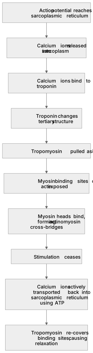
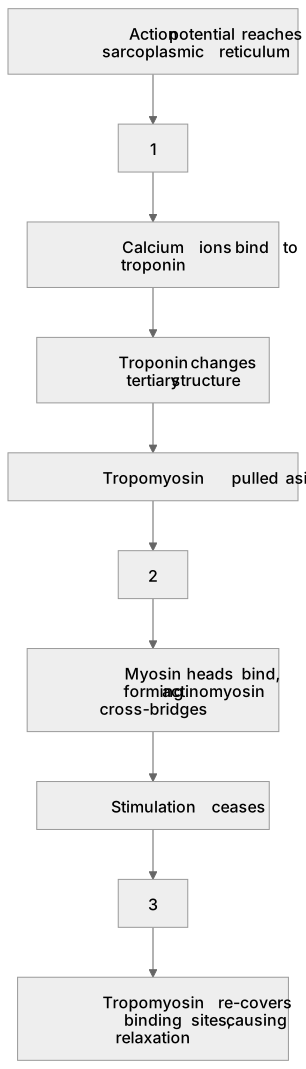
In relaxed muscle, tropomyosin covers myosin binding sites on actin filaments, preventing actinomyosin bridge formation. When an action potential reaches the sarcoplasmic reticulum, it releases calcium ions into the sarcoplasm. These ions bind to troponin, altering its tertiary structure. This pulls tropomyosin aside, exposing the binding sites. Myosin heads attach, forming cross-bridges to initiate contraction. For example, during a sustained bicep curl, continuous calcium release maintains exposed sites for repeated power strokes. When stimulation ceases, calcium ions are actively transported back into the sarcoplasmic reticulum using ATP. Tropomyosin then re-covers the sites, causing relaxation.
What earns the marks
- Calcium ions diffuse from the sarcoplasmic reticulum into the sarcoplasm following an action potential.
- Calcium ions bind to troponin, causing it to change its tertiary structure.
- Myosin heads bind to the exposed sites on actin, forming actinomyosin cross-bridges.
- Muscle relaxation requires ATP to actively transport calcium ions back into the sarcoplasmic reticulum, allowing tropomyosin to block the sites again.
Common mistakes and how to put them right
Stating that calcium ions bind directly to tropomyosin.
Put it right: Calcium ions bind to troponin, which then undergoes a conformational change to move tropomyosin.
Confusing the roles of calcium ions and ATP in the bridge cycle.
Put it right: Calcium ions expose the binding sites, whereas ATP binding is required to break the actinomyosin cross-bridges.
Referring to the binding sites on actin as 'active sites'.
Put it right: Use the term 'binding sites', as 'active sites' strictly refers to enzymes.
Forgetting that calcium ion reuptake is an active process.
Put it right: Specify that calcium ions are actively transported back into the sarcoplasmic reticulum using ATP.
Exam tips
- If a question asks about factors affecting the force of muscle contraction, link this to the number of actinomyosin cross-bridges formed, which depends on calcium ion availability.
- Always specify the exact source and destination of calcium ions; state they are released from and actively pumped back into the sarcoplasmic reticulum, not just 'the cell'.
Try the lesson
The start of the Skeletal muscles are stimulated to contract by nerves and act as: The roles of calcium ions and tropomyosin in the cycle of actinomyosin bridge formation. lesson, made from checked teaching notes for this specification point.
Have a go first
What is the role of calcium ions in the cycle of actinomyosin bridge formation?
Listen
In relaxed muscle, tropomyosin covers myosin binding sites on actin filaments, preventing actinomyosin bridge formation. When an action potential reaches the sarcoplasmic reticulum, it releases calcium ions into the sarcoplasm. These ions bind to troponin, altering its tertiary structure. This pulls tropomyosin aside, exposing the binding sites. Myosin heads attach, forming cross-bridges to initiate contraction. For example, during a sustained bicep curl, continuous calcium release maintains exposed sites for repeated power strokes. When stimulation ceases, calcium ions are actively transported back into the sarcoplasmic reticulum using ATP. Tropomyosin then re-covers the sites, causing relaxation. A strong answer earns marks for these points. Calcium ions diffuse from the sarcoplasmic reticulum into the sarcoplasm following an action potential. Calcium ions bind to troponin, causing it to change its tertiary structure. Myosin heads bind to the exposed sites on actin, forming actinomyosin cross-bridges. Muscle relaxation requires ATP to actively transport calcium ions back into the sarcoplasmic reticulum, allowing tropomyosin to block the sites again. A common slip: Stating that calcium ions bind directly to tropomyosin. Instead, Calcium ions bind to troponin, which then undergoes a conformational change to move tropomyosin. Exam tip: If a question asks about factors affecting the force of muscle contraction, link this to the number of actinomyosin cross-bridges formed, which depends on calcium ion availability.
See it
Key words
Say what each word means before you turn the card.
Tropomyosin and troponin function
Try answering aloud or in your head before turning the card.
Calcium ion release and active reuptake
Try answering aloud or in your head before turning the card.
Actinomyosin cross-bridge formation
Try answering aloud or in your head before turning the card.
See every definition
- Tropomyosin and troponin function
- In relaxed muscle, tropomyosin covers myosin binding sites on actin filaments; calcium ions bind to troponin, altering its tertiary structure and pulling tropomyosin aside to expose the binding sites.
- Calcium ion release and active reuptake
- An action potential reaching the sarcoplasmic reticulum releases calcium ions into the sarcoplasm; when stimulation ceases, calcium ions are actively transported back into the sarcoplasmic reticulum using ATP.
- Actinomyosin cross-bridge formation
- Myosin heads attach to the exposed binding sites on actin, forming cross-bridges to initiate contraction.
Label the diagram
Some boxes have lost their names. Pick the right name for each number.
1Box 1
2Box 2
3Box 3
There is more to this lesson
The full lesson goes on to check your understanding and help you earn the marks. Create a free account to carry on, with your progress saved.
A week of Premium from your first lesson · Then the free plan continues · No card, no charge
Test your understanding of Skeletal muscles are stimulated to contract by nerves and act as: The roles of calcium ions and tropomyosin in the cycle of actinomyosin bridge formation.
Active recall beats re-reading. Take an instant quiz now to test what you’ve learned and lock it into long-term memory.
A week of Premium from your first lesson · Then the free plan continues · No card, no charge