OCR · A-Level · Biology
Course version: H420 · Version from Sept 2025
(a) the use of microscopy to observe and investigate different types of…
Full specification wording
Cell structure: (a) the use of microscopy to observe and investigate different types of cell and cell structure in a range of eukaryotic organisms
Microscopy lets you observe and investigate cell structure across many eukaryotic organisms.
Checked against the OCR H420 specification (opens in a new tab) on .
- Point 1 of 10
- 4 key terms
Study notes
From the checked teaching notes for this specification point.
The idea
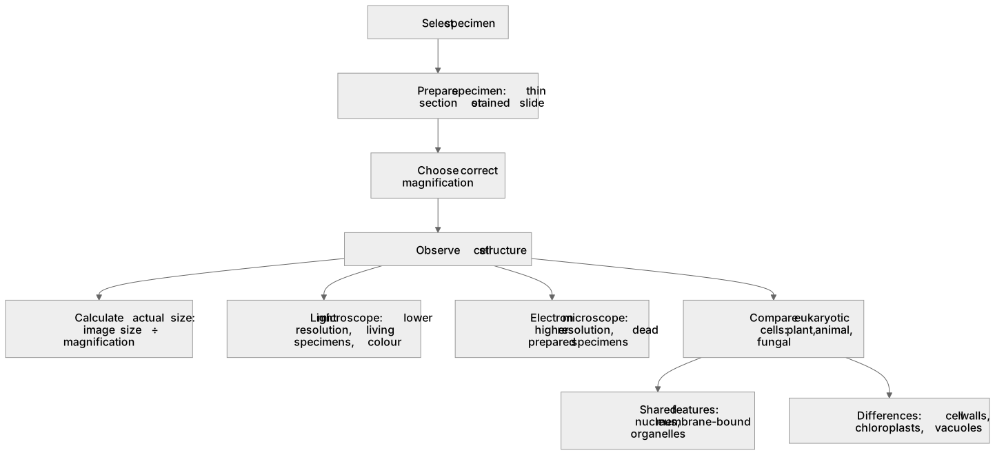
Microscopy lets you observe and investigate cell structure across many eukaryotic organisms. Light microscopes use visible light and give lower magnification and resolution but allow living specimens and colour; electron microscopes use a beam of electrons, giving far higher resolution and revealing organelles such as mitochondria and ribosomes, but require dead, prepared specimens. To investigate, you select a specimen, prepare it (for example a thin section or a stained slide), choose the correct magnification, and calculate actual size using magnification = image size ÷ actual size. Comparing cells from different eukaryotes, such as plant, animal and fungal cells, shows shared features (nucleus, membrane-bound organelles) and differences (cell walls, chloroplasts, vacuoles).
What earns the marks
- Light microscopes have lower resolution than electron microscopes but can view living specimens and show colour.
- Electron microscopes give higher magnification and resolution, revealing organelles such as mitochondria and ribosomes, but specimens are dead.
- Magnification is calculated as image size divided by actual size, allowing real cell dimensions to be determined.
- Comparing eukaryotic cells (plant, animal, fungal) reveals shared organelles and structural differences such as cell walls and chloroplasts.
- Correct specimen preparation and choice of magnification are needed to observe cell structure clearly.
Common mistakes and how to put them right
Confusing magnification with resolution.
Put it right: Magnification enlarges the image, while resolution is the ability to distinguish two close points.
Thinking electron microscopes can view living cells.
Put it right: Specimens must be dead and prepared in a vacuum.
Using the magnification formula the wrong way round.
Put it right: Actual size = image size ÷ magnification.
Exam tips
- State clearly whether a feature is seen with a light or electron microscope when comparing cell structure.
- Show the magnification calculation with correct units, converting to micrometres where needed.
- Link observed structures to the type of eukaryotic organism, for example chloroplasts in plant cells.
Try the lesson
The start of the Cell structure: (a) the use of microscopy to observe and investigate different types of cell and cell structure in a range of eukaryotic organisms lesson, made from checked teaching notes for this specification point.
Have a go first
A student wants to see the internal details of a mitochondrion in a eukaryotic cell. Which type of microscope is most suitable?
Listen
Microscopy lets you observe and investigate cell structure across many eukaryotic organisms. Light microscopes use visible light and give lower magnification and resolution but allow living specimens and colour; electron microscopes use a beam of electrons, giving far higher resolution and revealing organelles such as mitochondria and ribosomes, but require dead, prepared specimens. To investigate, you select a specimen, prepare it (for example a thin section or a stained slide), choose the correct magnification, and calculate actual size using magnification = image size ÷ actual size. Comparing cells from different eukaryotes, such as plant, animal and fungal cells, shows shared features (nucleus, membrane-bound organelles) and differences (cell walls, chloroplasts, vacuoles). A strong answer earns marks for these points. Light microscopes have lower resolution than electron microscopes but can view living specimens and show colour. Electron microscopes give higher magnification and resolution, revealing organelles such as mitochondria and ribosomes, but specimens are dead. Magnification is calculated as image size divided by actual size, allowing real cell dimensions to be determined. Comparing eukaryotic cells (plant, animal, fungal) reveals shared organelles and structural differences such as cell walls and chloroplasts. A common slip: Confusing magnification with resolution. Instead, magnification enlarges the image, while resolution is the ability to distinguish two close points. Exam tip: State clearly whether a feature is seen with a light or electron microscope when comparing cell structure.
See it
Watch it explained
What is the one idea the video explains? Say it in a sentence.
Read the described scene instead
Microscopy is how biologists observe and investigate cell structure across a wide range of eukaryotic organisms. Every cell we discuss here is observed through a microscope. So before comparing cells, we need to know what each instrument can and cannot show. Light microscopes use visible light. They give lower magnification and resolution, but you can view living specimens and see colour. Electron microscopes use a beam of electrons. They give far higher magnification and resolution, revealing organelles such as mitochondria and ribosomes, but the specimen must be dead and prepared in a vacuum. To investigate a specimen, you select it, then prepare it — for example a thin section or a stained slide. Next you choose the correct magnification, and observe. Correct preparation and the right magnification are what let you see cell structure clearly. Now a calculation. Magnification equals image size divided by actual size. Rearranged, actual size equals image size divided by magnification. Suppose an image measures fifty millimetres and the magnification is five hundred. Pause here and work out the actual size. Dividing fifty by five hundred gives nought point one millimetres, which is one hundred micrometres. Always state the units, converting to micrometres where needed. Three traps to avoid. Magnification enlarges the image; resolution is the ability to distinguish two close points — they are not the same thing. Electron microscopes cannot view living cells, because specimens must be dead and prepared in a vacuum. And don't turn the formula upside down: actual size equals image size divided by magnification. Comparing cells from different eukaryotes shows what is shared and what differs. Plant, animal and fungal cells all have a nucleus and membrane-bound organelles. Differences include cell walls, chloroplasts and vacuoles. Link each structure you observe to the type of organism it came from. So: light microscopy for living, coloured specimens at lower resolution; electron microscopy for high-resolution detail in dead, prepared specimens. Prepare, magnify, calculate with image size divided by magnification, and compare structures across plant, animal and fungal cells.
Key words
Say what each word means before you turn the card.
Light versus electron microscopy
Try answering aloud or in your head before turning the card.
Calculating magnification and actual cell size
Try answering aloud or in your head before turning the card.
Comparing cell structure across eukaryotic organisms
Try answering aloud or in your head before turning the card.
Specimen preparation and observation of organelles
Try answering aloud or in your head before turning the card.
See every definition
- Light versus electron microscopy
- Light microscopes use visible light and give lower magnification and resolution but allow living specimens and colour, while electron microscopes use a beam of electrons and give far higher resolution.
- Calculating magnification and actual cell size
- Actual size is found using magnification = image size ÷ actual size, allowing real cell dimensions to be determined.
- Comparing cell structure across eukaryotic organisms
- Plant, animal and fungal cells show shared features such as a nucleus and membrane-bound organelles, and differences such as cell walls, chloroplasts and vacuoles.
- Specimen preparation and observation of organelles
- A specimen is selected and prepared, for example as a thin section or a stained slide, and the correct magnification is chosen to observe cell structure clearly.
Label the diagram
Some boxes have lost their names. Pick the right name for each number.
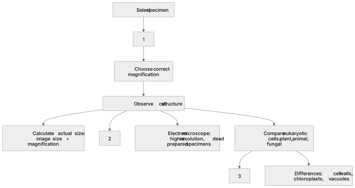
1Box 1
2Box 2
3Box 3
There is more to this lesson
The full lesson goes on to check your understanding and help you earn the marks. Create a free account to carry on, with your progress saved.
A week of Premium from your first lesson · Then the free plan continues · No card, no charge
Test your understanding of Cell structure: (a) the use of microscopy to observe and investigate different types of cell and cell structure in a range of eukaryotic organisms
Active recall beats re-reading. Take an instant quiz now to test what you’ve learned and lock it into long-term memory.
A week of Premium from your first lesson · Then the free plan continues · No card, no charge