OCR · A-Level · Biology
Course version: H420 · Version from Sept 2025
Cell structure: (c) the use of staining in light microscopy
This point requires learners to understand why stains are used in light microscopy and to know named examples.
Checked against the OCR H420 specification (opens in a new tab) on .
- Point 3 of 10
- 4 key terms
Study notes
From the checked teaching notes for this specification point.
The idea
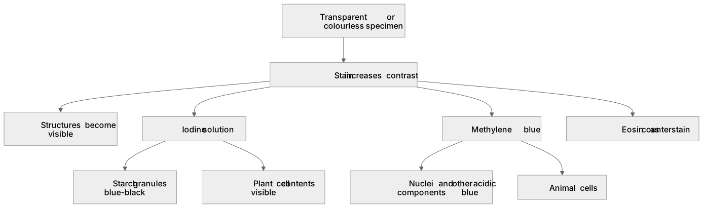
This point requires learners to understand why stains are used in light microscopy and to know named examples. Staining increases contrast because many biological specimens are transparent and have little natural colour, so without a stain cell structures are hard to distinguish. Different stains bind to different components: iodine solution stains starch granules blue-black and is used to make plant cell contents visible; methylene blue stains nuclei and other acidic components blue in animal cells; eosin can be used as a counterstain. Learners should be able to select the appropriate stain for a named structure and explain the resulting colour change. MCQ items may ask why a stain is needed, which stain is suitable for a particular specimen, or what colour a structure appears after staining.
What earns the marks
- Stains increase contrast so that transparent or colourless cell structures become visible.
- Iodine solution stains starch blue-black and is used with plant tissue.
- Methylene blue stains nuclei and other acidic components blue in animal cells.
- The choice of stain depends on the structure or molecule to be highlighted.
Common mistakes and how to put them right
Learners think stains magnify the specimen.
Put it right: Stains increase contrast, while magnification is provided by the lenses.
Learners assume one stain works for all structures.
Put it right: Different stains bind to different components.
Learners confuse the colour produced by iodine on starch.
Put it right: Starch stains blue-black.
Exam tips
- Link each named stain to the structure it highlights and the colour produced.
- When asked why a stain is used, focus on contrast and visibility rather than magnification.
- Check whether the specimen is plant or animal before choosing a stain.
Try the lesson
The start of the Cell structure: (c) the use of staining in light microscopy lesson, made from checked teaching notes for this specification point.
Have a go first
What is the main benefit of using a stain when examining an unstained specimen under a light microscope?
Listen
This point requires learners to understand why stains are used in light microscopy and to know named examples. Staining increases contrast because many biological specimens are transparent and have little natural colour, so without a stain cell structures are hard to distinguish. Different stains bind to different components: iodine solution stains starch granules blue-black and is used to make plant cell contents visible; methylene blue stains nuclei and other acidic components blue in animal cells; eosin can be used as a counterstain. Learners should be able to select the appropriate stain for a named structure and explain the resulting colour change. MCQ items may ask why a stain is needed, which stain is suitable for a particular specimen, or what colour a structure appears after staining. A strong answer earns marks for these points. Stains increase contrast so that transparent or colourless cell structures become visible. Iodine solution stains starch blue-black and is used with plant tissue. Methylene blue stains nuclei and other acidic components blue in animal cells. The choice of stain depends on the structure or molecule to be highlighted. A common slip: Learners think stains magnify the specimen. Instead, stains increase contrast, while magnification is provided by the lenses. Exam tip: Link each named stain to the structure it highlights and the colour produced.
See it
Key words
Say what each word means before you turn the card.
Purpose of staining
Try answering aloud or in your head before turning the card.
Named stains and their targets
Try answering aloud or in your head before turning the card.
Colour changes observed
Try answering aloud or in your head before turning the card.
Contrast versus magnification
Try answering aloud or in your head before turning the card.
See every definition
- Purpose of staining
- Staining increases contrast because many biological specimens are transparent and have little natural colour, so without a stain cell structures are hard to distinguish.
- Named stains and their targets
- Iodine solution stains starch granules and is used to make plant cell contents visible, while methylene blue stains nuclei and other acidic components in animal cells.
- Colour changes observed
- Iodine solution stains starch blue-black, and methylene blue stains nuclei and other acidic components blue.
- Contrast versus magnification
- Stains increase contrast, while magnification is provided by the lenses.
Label the diagram
Some boxes have lost their names. Pick the right name for each number.
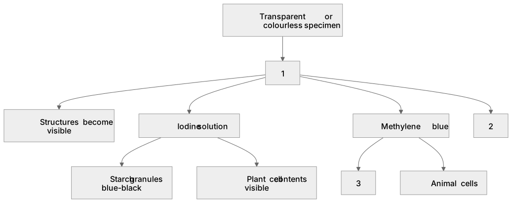
1Box 1
2Box 2
3Box 3
There is more to this lesson
The full lesson goes on to check your understanding and help you earn the marks. Create a free account to carry on, with your progress saved.
A week of Premium from your first lesson · Then the free plan continues · No card, no charge
Test your understanding of Cell structure: (c) the use of staining in light microscopy
Active recall beats re-reading. Take an instant quiz now to test what you’ve learned and lock it into long-term memory.
A week of Premium from your first lesson · Then the free plan continues · No card, no charge