OCR · A-Level · Biology
Course version: H420 · Version from Sept 2025
(l) (i) the structure of mammalian muscle and the mechanism of muscular…
Full specification wording
Plant and animal responses: (l) (i) the structure of mammalian muscle and the mechanism of muscular contraction
Mammalian skeletal muscle is made of bundles of fibres containing myofibrils.
Checked against the OCR H420 specification (opens in a new tab) on .
- Point 11 of 12
- 4 key terms
Study notes
From the checked teaching notes for this specification point.
The idea
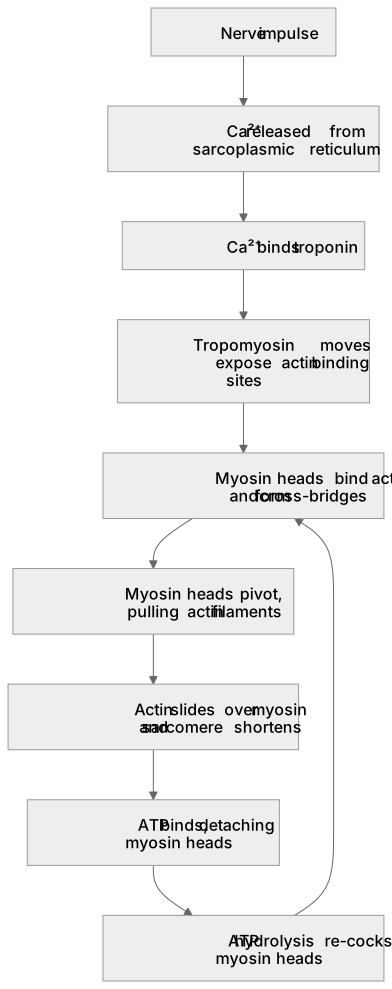
Mammalian skeletal muscle is made of bundles of fibres containing myofibrils. Each myofibril shows repeating sarcomeres bounded by Z-lines, with thick myosin filaments and thin actin filaments. The sliding filament model explains contraction: a nerve impulse releases calcium ions from the sarcoplasmic reticulum; Ca²⁺ binds troponin, moving tropomyosin to expose actin binding sites; myosin heads bind actin, form cross-bridges and pivot, pulling actin filaments towards the centre of the sarcomere. ATP binding detaches myosin heads and ATP hydrolysis re-cocks them for the next cycle. As actin slides over myosin, the sarcomere shortens and the muscle contracts, while the filaments themselves do not change length.
What earns the marks
- Skeletal muscle fibres contain myofibrils with repeating sarcomeres bounded by Z-lines, containing actin and myosin filaments.
- A nerve impulse triggers release of Ca²⁺ from the sarcoplasmic reticulum.
- Ca²⁺ binds troponin, moving tropomyosin to expose actin binding sites.
- Myosin heads bind actin to form cross-bridges and pivot, sliding actin filaments and shortening the sarcomere.
- ATP is required to detach myosin heads and to re-cock them for further cycles.
Common mistakes and how to put them right
Thinking the filaments themselves shorten.
Put it right: The filaments slide past each other and the sarcomere shortens, not the actin or myosin.
Omitting the role of Ca²⁺ and troponin.
Put it right: Without Ca²⁺ binding troponin, tropomyosin blocks the binding sites and cross-bridges cannot form.
Forgetting ATP is needed for detachment as well as contraction.
Put it right: Without ATP, myosin heads cannot release actin.
Exam tips
- Sequence the events in order: impulse, Ca²⁺ release, troponin/tropomyosin, cross-bridge, power stroke, ATP detachment.
- Name the structures precisely (sarcomere, Z-line, actin, myosin, sarcoplasmic reticulum) to gain credit.
- State clearly that the sarcomere shortens because filaments slide, not because filaments contract.
Try the lesson
The start of the Plant and animal responses: (l) (i) the structure of mammalian muscle and the mechanism of muscular contraction lesson, made from checked teaching notes for this specification point.
Have a go first
What is the functional unit of contraction in a skeletal muscle fibre called?
Listen
Mammalian skeletal muscle is made of bundles of fibres containing myofibrils. Each myofibril shows repeating sarcomeres bounded by Z-lines, with thick myosin filaments and thin actin filaments. The sliding filament model explains contraction: a nerve impulse releases calcium ions from the sarcoplasmic reticulum; Ca²⁺ binds troponin, moving tropomyosin to expose actin binding sites; myosin heads bind actin, form cross-bridges and pivot, pulling actin filaments towards the centre of the sarcomere. ATP binding detaches myosin heads and ATP hydrolysis re-cocks them for the next cycle. As actin slides over myosin, the sarcomere shortens and the muscle contracts, while the filaments themselves do not change length. A strong answer earns marks for these points. Skeletal muscle fibres contain myofibrils with repeating sarcomeres bounded by Z-lines, containing actin and myosin filaments. A nerve impulse triggers release of Ca²⁺ from the sarcoplasmic reticulum. Ca²⁺ binds troponin, moving tropomyosin to expose actin binding sites. Myosin heads bind actin to form cross-bridges and pivot, sliding actin filaments and shortening the sarcomere. Exam tip: Sequence the events in order: impulse, Ca²⁺ release, troponin/tropomyosin, cross-bridge, power stroke, ATP detachment.
See it
Key words
Say what each word means before you turn the card.
Sarcomere structure
Try answering aloud or in your head before turning the card.
Sliding filament model
Try answering aloud or in your head before turning the card.
Role of Ca²⁺ and troponin
Try answering aloud or in your head before turning the card.
ATP in the cross-bridge cycle
Try answering aloud or in your head before turning the card.
See every definition
- Sarcomere structure
- Repeating units of a myofibril bounded by Z-lines, containing thick myosin filaments and thin actin filaments.
- Sliding filament model
- Actin filaments slide over myosin as myosin heads pivot, so the sarcomere shortens while the filaments themselves do not change length.
- Role of Ca²⁺ and troponin
- Calcium ions released from the sarcoplasmic reticulum bind troponin, moving tropomyosin to expose actin binding sites.
- ATP in the cross-bridge cycle
- ATP binding detaches myosin heads from actin and ATP hydrolysis re-cocks them for the next cycle.
Label the diagram
Some boxes have lost their names. Pick the right name for each number.
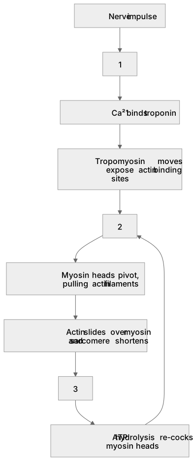
1Box 1
2Box 2
3Box 3
There is more to this lesson
The full lesson goes on to check your understanding and help you earn the marks. Create a free account to carry on, with your progress saved.
A week of Premium from your first lesson · Then the free plan continues · No card, no charge
Test your understanding of Plant and animal responses: (l) (i) the structure of mammalian muscle and the mechanism of muscular contraction
Active recall beats re-reading. Take an instant quiz now to test what you’ve learned and lock it into long-term memory.
A week of Premium from your first lesson · Then the free plan continues · No card, no charge