Study Notes
Overview
Welcome to the ultimate guide on Amino Acids, Proteins, and DNA. Although often categorised under biological chemistry, this topic is a rigorous, deeply chemical exploration of the molecules that build and sustain life. You are not just learning biology here; you are applying core organic chemistry principles—such as acid-base equilibria, condensation reactions, hydrogen bonding, and stereochemistry—to complex biological macromolecules.
Why is this important for your exams? This topic serves as a critical bridge. It links your foundational knowledge of functional groups and intermolecular forces to real-world applications, including enzyme catalysis and the precise mechanism of anti-cancer drugs like cisplatin. Examiners frequently use this section to test your ability to synthesize knowledge across the specification. Expect questions that ask you to draw zwitterions under different pH conditions, explain the stereospecificity of enzymes, or deduce the structure of a peptide chain from its constituent amino acids.
Mastering this topic requires moving beyond rote memorisation. You must understand why these molecules behave the way they do in different environments. This guide will provide the clear explanations, examiner commentary, and active recall strategies you need to secure top marks.
Level note: The supplied specification reference, AQA 3.3.13, is A-level-only content. There is no GCSE Foundation/Higher tier split for this topic; all material in this guide is A-level extension content. The chemistry has been introduced clearly so that a GCSE student can use it as a bridge to post-16 study.
Key Concepts
Concept 1: Amino Acids and Zwitterions
Alpha-amino acids are the fundamental building blocks of proteins. They are bifunctional compounds, containing both a basic amine group (-\text{NH}_2) and an acidic carboxyl group (-\text{COOH}) attached to the same central carbon atom (the alpha carbon). Because they contain both acidic and basic groups, they are amphoteric—they can react with both acids and bases.
In the solid state and in neutral aqueous solutions, amino acids do not exist as uncharged molecules. Instead, the acidic carboxyl group donates a proton to the basic amine group. This internal acid-base reaction forms a zwitterion, a dipolar ion that has both a positive charge (-\text{NH}_3^+) and a negative charge (-\text{COO}^-), but no overall net electrical charge.
The formation of zwitterions explains why amino acids have surprisingly high melting points; they are held together by strong electrostatic ionic bonds in a giant ionic lattice, rather than weak intermolecular forces.
Behaviour in Different pH EnvironmentsExaminers love to test your ability to predict the structure of an amino acid when the pH changes:
- In Acidic Solution (Low pH): There is an abundance of \text{H}^+ ions. The weakly basic -\text{COO}^- group accepts a proton to become -\text{COOH}. The molecule now has an overall positive charge (-\text{NH}_3^+ and -\text{COOH}).
- In Alkaline Solution (High pH): There is an abundance of \text{OH}^- ions. The weakly acidic -\text{NH}_3^+ group donates a proton to the \text{OH}^- to form water, reverting to -\text{NH}_2. The molecule now has an overall negative charge (-\text{NH}_2 and -\text{COO}^-).
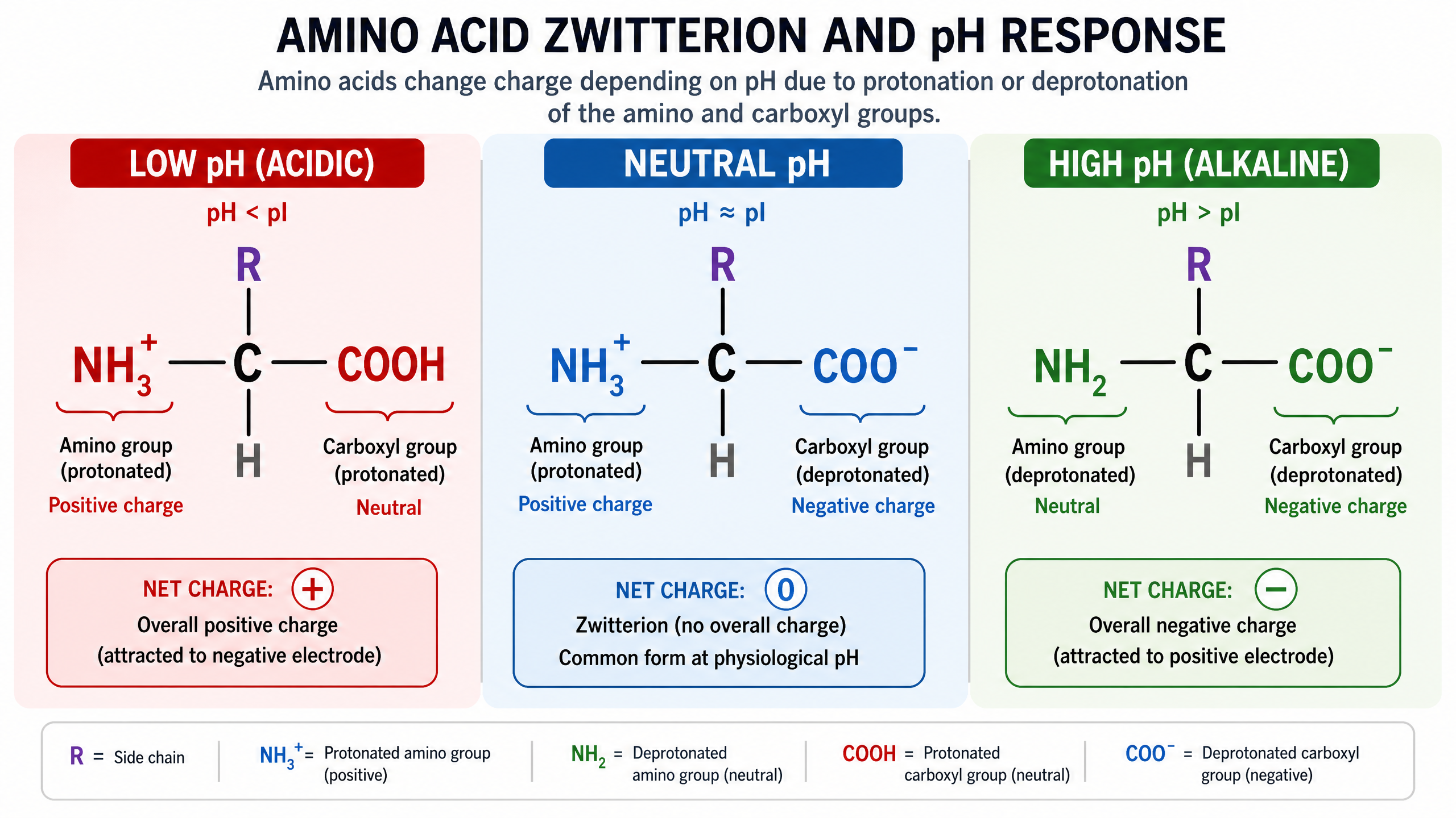
Example: Draw the structure of alanine (2-aminopropanoic acid) in a solution of pH 1.
Explanation: At pH 1, the solution is highly acidic. The zwitterion will accept a proton. The -\text{COO}^- group becomes -\text{COOH}, while the -\text{NH}_3^+ group remains protonated. The resulting ion is \text{CH}_3\text{CH}(\text{NH}_3^+)\text{COOH}.
Concept 2: Peptides and Proteins
Proteins are naturally occurring condensation polymers formed by joining together sequences of amino acids. The linkage that holds them together is an amide bond, specifically called a peptide link (-\text{CONH}-).
When two amino acids react, the carboxyl group of one molecule reacts with the amine group of the other. A molecule of water is eliminated (hence, a condensation reaction), and a dipeptide is formed. This process can continue to form tripeptides, polypeptides, and eventually massive protein chains.
Hydrolysis of ProteinsThe formation of a peptide link is reversible. Proteins and peptides can be broken back down into their constituent amino acids via hydrolysis. In the laboratory, this requires harsh conditions: boiling the protein with concentrated hydrochloric acid (\text{HCl}) for about 24 hours. In biological systems, enzymes perform this hydrolysis rapidly at body temperature.
Because the laboratory hydrolysis uses concentrated acid, the amino acids produced will be in their fully protonated, positively charged cationic form. Examiners will penalise you if you draw the neutral amino acid instead of the protonated form when describing acid hydrolysis.
Concept 3: Protein Structure and Bonding
The function of a protein is entirely dependent on its three-dimensional shape. This shape is determined by a hierarchy of structures, each stabilized by specific types of chemical bonds.
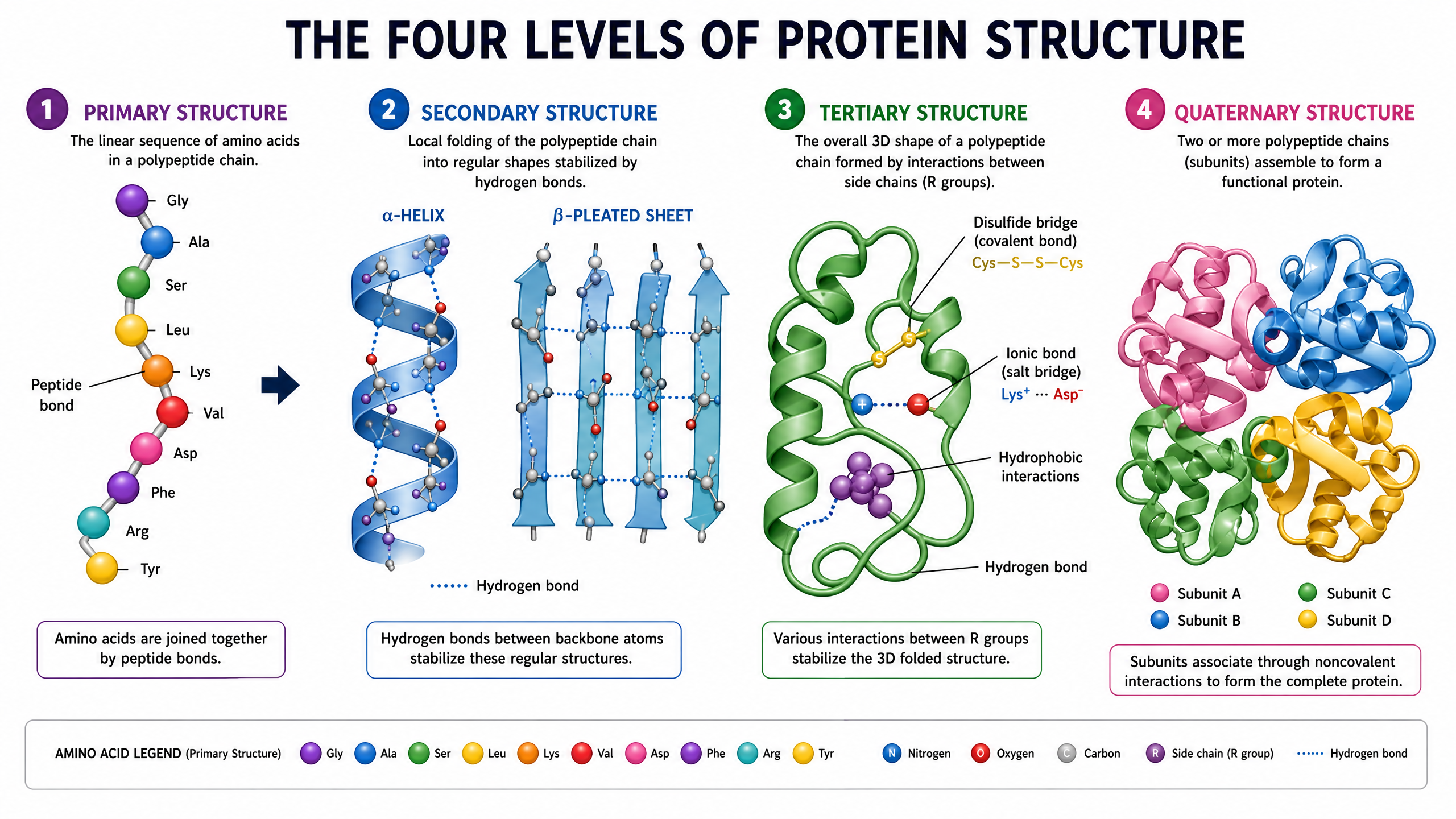
- Primary Structure: This is the linear sequence of amino acids in the polypeptide chain. It is held together entirely by strong, covalent peptide bonds. This sequence is determined by the genetic code in DNA.
- Secondary Structure: The polypeptide chain does not remain straight. It folds or coils into regular patterns, primarily the \alpha-helix and the \beta-pleated sheet. These structures are maintained by hydrogen bonds that form between the \text{C=O} group of one peptide link and the \text{N-H} group of another peptide link further along the chain. These hydrogen bonds are individually weak but collectively very strong.
- Tertiary Structure: This is the overall 3D folding of the entire protein molecule into a specific, complex shape. The tertiary structure is crucial for the function of enzymes and is maintained by several types of interactions between the 'R' groups (side chains) of the amino acids:
- Hydrogen bonds: Between polar R groups (e.g., those containing -\text{OH} or -\text{NH}_2).
- Ionic bonds: Between charged R groups (e.g., -\text{NH}_3^+ and -\text{COO}^-). These are highly sensitive to pH changes. If the pH changes, the charges on the R groups may be lost, breaking the ionic bonds and causing the protein to denature (lose its specific 3D shape).
- Disulfide bridges (Sulfur-sulfur bonds): These are strong covalent bonds that form between the sulfur atoms of two cysteine amino acids via an oxidation reaction. They act as molecular "safety pins," firmly holding different parts of the folded chain together. They are not broken by gentle heating or mild pH changes.
Concept 4: Enzymes and Stereospecificity
Enzymes are globular proteins that act as biological catalysts. They speed up chemical reactions by providing an alternative reaction pathway with a lower activation energy. They do this without being used up in the reaction.
The catalytic action occurs at a specific region on the enzyme called the active site. The active site is a 3D crevice or cleft formed by the tertiary structure of the protein. For a reaction to occur, the reacting molecule, known as the substrate, must fit perfectly into the active site, much like a key fitting into a lock.
Because the active site has a very precise 3D shape defined by chiral amino acids, enzymes are highly stereospecific. If a substrate exists as a pair of enantiomers (optical isomers), the active site will typically only accommodate one of the enantiomers. The other enantiomer will have the wrong spatial arrangement of groups and will not fit, meaning the enzyme cannot catalyse its reaction. This is a common application of stereochemistry in exam questions.
Enzyme InhibitionDrugs can be designed to act as enzyme inhibitors. An inhibitor is a molecule that has a similar shape to the natural substrate. It can bind to the active site, blocking the actual substrate from entering. If the substrate cannot bind, the enzyme-catalysed reaction cannot proceed. Computers are heavily used in modern drug design to model the 3D shape of active sites and design perfectly fitting inhibitor molecules, reducing the need for trial-and-error synthesis in the laboratory.
Concept 5: DNA Structure
Deoxyribonucleic acid (DNA) is the molecule that stores the genetic instructions for life. It is a condensation polymer formed from monomers called nucleotides.
Every nucleotide consists of three components:
- A phosphate ion.
- A pentose sugar called 2-deoxyribose.
- A nitrogenous base (Adenine, Cytosine, Guanine, or Thymine). The structures of these bases are provided in the Chemistry Data Booklet; you do not need to memorise them, but you must know how to use them.
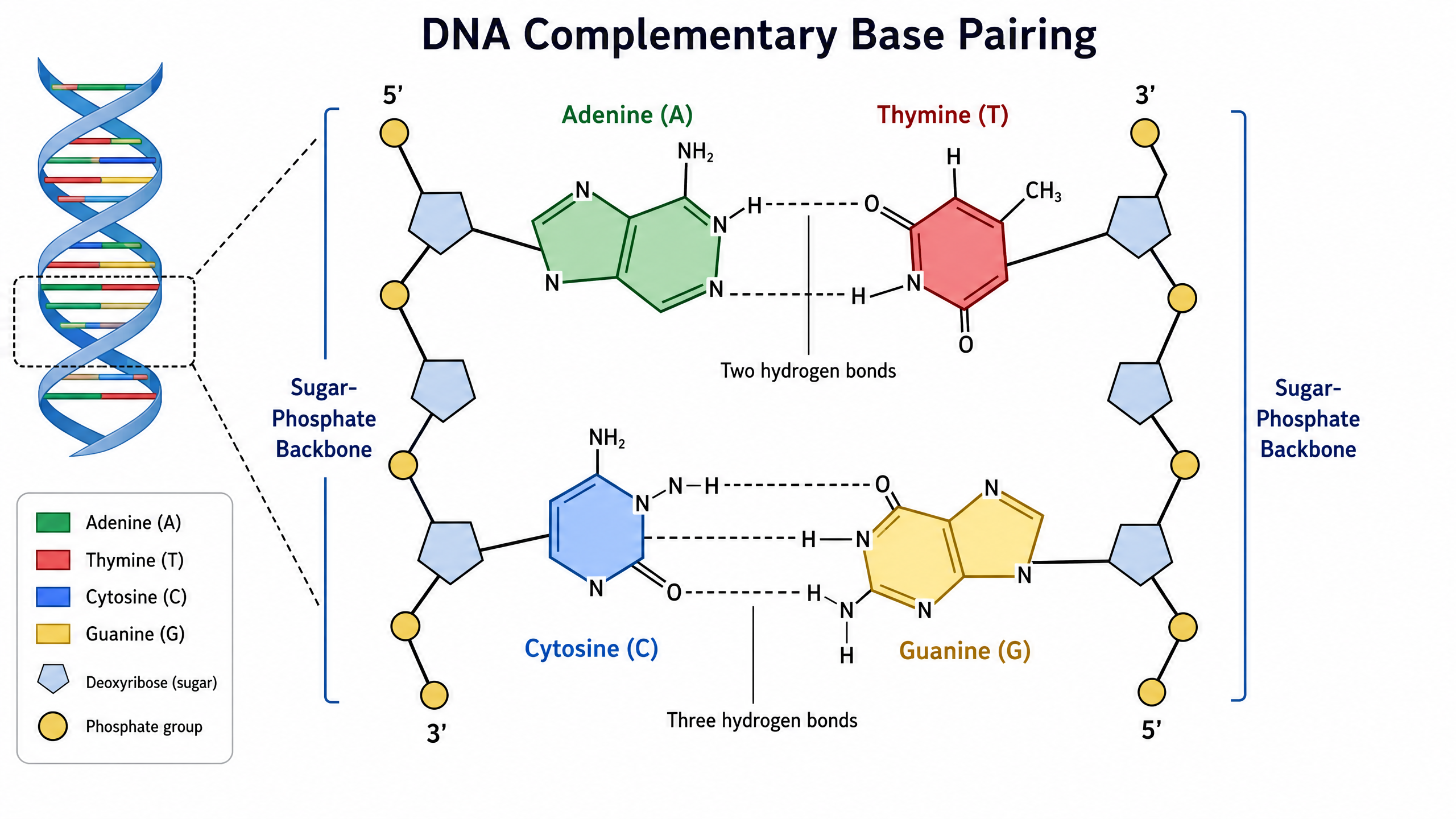
The nucleotides join together via condensation reactions. A covalent bond forms between the phosphate group of one nucleotide and the 2-deoxyribose sugar of the next. This creates a long, continuous sugar-phosphate backbone, with the bases protruding from the sugars.
DNA exists as two complementary strands that wind around each other to form a double helix. The two strands are held together by hydrogen bonding between specific pairs of bases:
- Adenine (A) always pairs with Thymine (T) via two hydrogen bonds.
- Cytosine (C) always pairs with Guanine (G) via three hydrogen bonds.
This specific base pairing is the chemical basis for DNA replication and the accurate transmission of genetic information. When cells divide, the hydrogen bonds break, the strands separate, and new complementary strands are built along each template.
Concept 6: The Action of Cisplatin
Cisplatin is a highly effective anti-cancer drug. Its chemical name is cis-diamminedichloroplatinum(II), and its formula is [\text{Pt}(\text{NH}_3)_2\text{Cl}_2]. It is a square planar complex of platinum(II). The two chloride ligands and the two ammonia ligands are arranged adjacent to each other (the cis isomer).
Cancer involves the uncontrolled division and replication of cells. Cisplatin works by stopping DNA replication in these rapidly dividing cells.
**Mechanism of Action:**Inside the cell, cisplatin undergoes a ligand replacement reaction. The chloride ligands are displaced by water, and then a bond forms between the platinum atom and a nitrogen atom on a guanine base within the DNA double helix. A second nitrogen atom on an adjacent guanine base replaces the other chloride ligand. This cross-linking causes the DNA strand to kink or distort. The distortion prevents the enzymes responsible for DNA replication from moving along the strand, thereby stopping the cancer cell from dividing.
**Adverse Effects:**The major drawback of cisplatin is that it is not perfectly selective. It binds to the DNA of any rapidly dividing cell, not just cancer cells. This leads to severe side effects, such as hair loss, nausea, and immune suppression (due to the drug affecting hair follicles, the stomach lining, and white blood cells). Society and medical professionals must constantly assess the balance between the life-saving benefits of such drugs and their harsh adverse effects.
Mathematical/Scientific Relationships
-
**Retention Factor (R_f) in Chromatography — Must memorise:**When separating amino acids using thin-layer chromatography (TLC), you must identify them by calculating their R_f values. There are no other quantitative formulae specified for AQA 3.3.13, and no unit conversion is required because R_f has no units.
R_f = \frac{\text{Distance moved by the amino acid spot}}{\text{Distance moved by the solvent front}}
- The distance is measured from the original pencil baseline.
- R_f values have no units and are always less than 1.
- Because amino acids are colourless, the chromatogram must be sprayed with a developing agent, such as ninhydrin, or viewed under ultraviolet (UV) light to make the spots visible.
Practical Applications
Chromatography of Amino AcidsWhile there is no mandatory required practical specifically for amino acids, the technique of Thin-Layer Chromatography (TLC) is heavily tested in this context. Examiners frequently ask you to explain the steps and justify the precautions taken.
- Draw a pencil line near the bottom of a TLC plate. (Pencil is used because ink would dissolve in the solvent and interfere with the chromatogram).
- Place a small spot of the amino acid mixture on the line using a capillary tube.
- Place the plate in a beaker containing a shallow layer of solvent (the solvent level must be below the pencil line, otherwise the amino acids will dissolve into the bulk solvent rather than moving up the plate).
- Cover the beaker with a lid to prevent the toxic or flammable solvent from evaporating and to saturate the atmosphere inside the beaker.
- Allow the solvent to run up the plate until it is near the top.
- Remove the plate and immediately mark the solvent front with a pencil before it evaporates.
- Allow the plate to dry in a fume cupboard (as the solvent is likely toxic).
- Spray with ninhydrin in a fume cupboard. The amino acids will appear as purple/brown spots.
- Calculate the R_f values and compare them to a database of known values to identify the amino acids present. Note that R_f values depend on the specific solvent and temperature used.
Examiner Practical Focus: TLC of Amino Acids
Required-practical status: Amino-acid TLC is not a named AQA A-level Chemistry required practical within 3.3.13, but the technique and its data treatment are highly examinable. An appropriate apparatus list is: a TLC plate, capillary spotting tube, pencil, ruler, developing beaker with lid, suitable solvent, fume cupboard, UV lamp or ninhydrin, gloves and eye protection. Expected results are separated coloured spots after ninhydrin treatment; each spot has an R_f value below 1. Candidates gain credit for comparing values only where the solvent and conditions are the same.
Common errors include placing the baseline below the solvent level, using ink rather than pencil, failing to mark the solvent front immediately, making a spot too large, and measuring from the wrong point. Examiners test this by asking candidates to justify the pencil baseline, solvent level, lidded beaker, fume-cupboard use, and ninhydrin treatment. They also ask for R_f calculations, identification from standards, and a conclusion based on matching values. Graph skills are not normally required here; for data interpretation, use a table of spot distances and calculate each unitless R_f value carefully.
Visual Resources
3 diagrams and illustrations
Interactive Diagrams
2 interactive diagrams to visualise key concepts
Conceptual Flow Outline
Flowchart showing the steps for identifying amino acids using Thin-Layer Chromatography (TLC).
Conceptual Flow Outline
Process diagram illustrating the stereospecificity of enzyme action.
Worked Examples
3 detailed examples with solutions and examiner commentary
Practice Questions
Test your understanding — click to reveal model answers
Alanine has the formula CH3CH(NH2)COOH. Draw the structure of the dipeptide formed when two molecules of alanine react together. [2 marks]
Hint: Remember to remove a molecule of water (H2O) to form the peptide link (-CONH-).
A mixture of amino acids was separated by thin-layer chromatography. The solvent front moved 8.0 cm. A spot corresponding to the amino acid leucine moved 4.8 cm. Calculate the Rf value for leucine. [1 mark]
Hint: Rf = distance moved by spot / distance moved by solvent front.
Explain why the melting point of an amino acid is much higher than that of a carboxylic acid of similar relative molecular mass. [3 marks]
Hint: Think about the form amino acids take in the solid state.
A section of a protein chain contains the amino acid cysteine, which has a side chain containing a -SH group. Explain how this side chain contributes to the tertiary structure of the protein. [2 marks]
Hint: What type of bond forms between two sulfur atoms?
Explain why cisplatin is administered as a single pure enantiomer rather than a racemic mixture. [2 marks]
Hint: Think about stereospecificity and how the drug interacts with DNA.