AQA · A-Level · Physics
Course version: 7408 · Version from Sept 2016
Advantages of FTP detector compared with photographic detection.
Full specification wording
Image detection and enhancement (A-level only): Advantages of FTP detector compared with photographic detection.
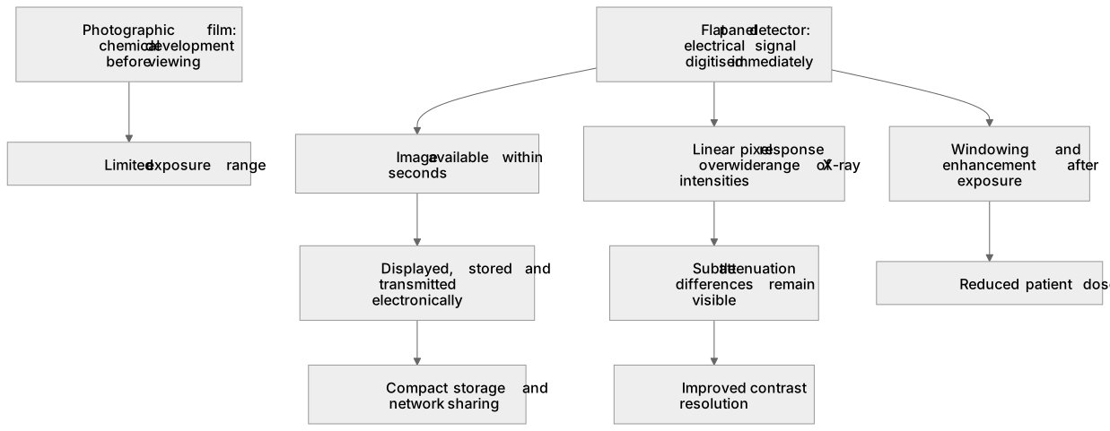
Photographic detection uses film that must be chemically developed before the image can be seen, and the film responds over a limited exposure range.
Checked against the AQA 7408 specification (opens in a new tab) on .
- Point 2 of 4
- 4 key terms
Study notes
From the checked teaching notes for this specification point.
The idea
Photographic detection uses film that must be chemically developed before the image can be seen, and the film responds over a limited exposure range. A flat panel detector instead produces an electrical signal that is digitised immediately, so the image is available within seconds and can be displayed, stored and transmitted electronically. Because the pixel response is linear over a wide range of X-ray intensities, subtle differences in attenuation remain visible, improving contrast resolution. The digital data can be windowed and enhanced after exposure without repeating the scan, reducing patient dose. There is no film to load, process, handle or archive physically, so workflow is faster and storage is compact. Electronic images can also be shared across networks for reporting and comparison with previous examinations.
What earns the marks
- Immediate electronic readout gives the image in seconds, whereas photographic film requires chemical development before viewing.
- Digital storage and transmission replace bulky film archives, allowing rapid access, copying and comparison with earlier images.
- A wide linear dynamic range means small differences in X-ray attenuation are recorded, improving contrast resolution compared with film.
- Post-acquisition processing such as windowing and enhancement can improve the displayed image without further patient exposure.
- Lower retake rates and reduced repeat exposures follow from immediate checking of image quality, lowering overall patient dose.
- No chemical developer or fixer is needed, removing processing consumables and associated waste.
Common mistakes and how to put them right
Claiming flat panel detectors use no radiation.
Put it right: They still require X-rays, but the dose per useful image can often be reduced.
Saying film gives an instant image.
Put it right: Film needs development time before it can be viewed.
Assuming digital images cannot be altered.
Put it right: They can be processed and enhanced, which is an advantage when used appropriately.
Exam tips
- Structure comparisons as paired points: what film does, what the flat panel detector does, and why that matters clinically.
- Use precise terms such as dynamic range, contrast resolution and post-acquisition processing rather than vague words like better.
- Where dose is mentioned, explain the mechanism, for example fewer repeat exposures because the image can be checked immediately.
Try the lesson
The start of the Image detection and enhancement (A-level only): Advantages of FTP detector compared with photographic detection. lesson, made from checked teaching notes for this specification point.
Have a go first
Which advantage does an FTP detector have over photographic film when a radiographer needs the image immediately?
Listen
Photographic detection uses film that must be chemically developed before the image can be seen, and the film responds over a limited exposure range. A flat panel detector instead produces an electrical signal that is digitised immediately, so the image is available within seconds and can be displayed, stored and transmitted electronically. Because the pixel response is linear over a wide range of X-ray intensities, subtle differences in attenuation remain visible, improving contrast resolution. The digital data can be windowed and enhanced after exposure without repeating the scan, reducing patient dose. There is no film to load, process, handle or archive physically, so workflow is faster and storage is compact. Electronic images can also be shared across networks for reporting and comparison with previous examinations. A strong answer earns marks for these points. Immediate electronic readout gives the image in seconds, whereas photographic film requires chemical development before viewing. Digital storage and transmission replace bulky film archives, allowing rapid access, copying and comparison with earlier images. A wide linear dynamic range means small differences in X-ray attenuation are recorded, improving contrast resolution compared with film. Post-acquisition processing such as windowing and enhancement can improve the displayed image without further patient exposure. A common slip: Claiming flat panel detectors use no radiation. Instead, they still require X-rays, but the dose per useful image can often be reduced. Exam tip: Structure comparisons as paired points: what film does, what the flat panel detector does, and why that matters clinically.
See it
Key words
Say what each word means before you turn the card.
Digital versus analogue imaging
Try answering aloud or in your head before turning the card.
Patient dose optimisation
Try answering aloud or in your head before turning the card.
Image quality and contrast resolution
Try answering aloud or in your head before turning the card.
Clinical workflow and data storage
Try answering aloud or in your head before turning the card.
See every definition
- Digital versus analogue imaging
- A flat panel detector produces an electrical signal that is digitised immediately, so the image is available within seconds, whereas photographic film must be chemically developed before the image can be seen.
- Patient dose optimisation
- Digital data can be windowed and enhanced after exposure without repeating the scan, and lower retake rates and reduced repeat exposures follow from immediate checking of image quality, lowering overall patient dose.
- Image quality and contrast resolution
- Because the pixel response is linear over a wide range of X-ray intensities, subtle differences in attenuation remain visible, improving contrast resolution.
- Clinical workflow and data storage
- There is no film to load, process, handle or archive physically, so workflow is faster and storage is compact, and electronic images can be shared across networks for reporting and comparison with previous examinations.
Label the diagram
Some boxes have lost their names. Pick the right name for each number.
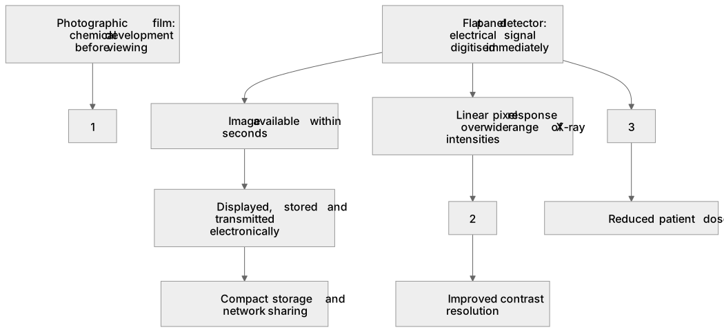
1Box 1
2Box 2
3Box 3
There is more to this lesson
The full lesson goes on to check your understanding and help you earn the marks. Create a free account to carry on, with your progress saved.
A week of Premium from your first lesson · Then the free plan continues · No card, no charge
Test your understanding of Image detection and enhancement (A-level only): Advantages of FTP detector compared with photographic detection.
Active recall beats re-reading. Take an instant quiz now to test what you’ve learned and lock it into long-term memory.
A week of Premium from your first lesson · Then the free plan continues · No card, no charge