AQA · A-Level · Physics
Course version: 7408 · Version from Sept 2016
Flat panel (FTP) detector including X-ray scintillator, photodiode pixels,…
Full specification wording
Image detection and enhancement (A-level only): Flat panel (FTP) detector including X-ray scintillator, photodiode pixels, electronic scanning.
A flat panel detector converts an X-ray shadow into a digital image.
Checked against the AQA 7408 specification (opens in a new tab) on .
- Point 1 of 4
- 4 key terms
Study notes
From the checked teaching notes for this specification point.
The idea
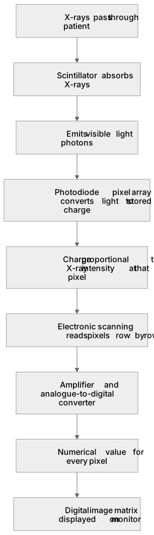
A flat panel detector converts an X-ray shadow into a digital image. X-rays passing through the patient strike a scintillator layer, commonly caesium iodide, which absorbs them and emits visible light photons. Beneath it lies a two-dimensional array of photodiode pixels, each a tiny light-sensitive semiconductor element that converts the light into an electrical charge stored locally. The charge at each pixel is therefore proportional to the X-ray intensity reaching that point. Readout electronics scan the array row by row, transferring each pixel's charge to an amplifier and analogue-to-digital converter, producing a numerical value for every pixel. These values form the digital image matrix displayed on a monitor. Scanning is rapid and automatic, so the image appears almost immediately after exposure.
What earns the marks
- The scintillator absorbs incident X-rays and re-emits visible light photons, acting as a converter between X-ray and optical energy.
- The photodiode array is a two-dimensional grid of pixels; each pixel converts received light into a stored electrical charge.
- Charge accumulated at a pixel is proportional to the X-ray intensity incident on that pixel, so spatial variation in X-ray transmission is encoded as charge variation.
- Electronic scanning reads pixels sequentially, typically row by row, using addressing circuitry rather than moving the detector.
- Signals pass through amplifiers and analogue-to-digital conversion so each pixel becomes a numerical grey level in a digital matrix.
- The resulting matrix is processed and displayed, giving an image whose contrast reflects differing X-ray attenuation within the patient.
Common mistakes and how to put them right
Thinking the scintillator directly produces the electrical signal.
Put it right: The scintillator emits visible light and the photodiode converts that light into charge.
Believing the whole panel is exposed and read simultaneously.
Put it right: Pixels are addressed and read out by electronic scanning, usually row by row.
Confusing the detector with photographic film by saying X-rays darken the panel.
Put it right: X-ray intensity is converted to light, then to charge, then to digital numbers.
Exam tips
- Draw a labelled layer diagram: scintillator on top, photodiode pixel array below, addressing and readout electronics at the edge.
- Use the chain X-ray to light to charge to digital number to display, naming the component responsible at each stage.
- Link pixel charge to X-ray attenuation in the patient so the answer explains image formation, not just hardware.
Try the lesson
The start of the Image detection and enhancement (A-level only): Flat panel (FTP) detector including X-ray scintillator, photodiode pixels, electronic scanning. lesson, made from checked teaching notes for this specification point.
Have a go first
In a flat panel (FTP) detector, which component converts the incoming X-ray photons into visible light?
Listen
A flat panel detector converts an X-ray shadow into a digital image. X-rays passing through the patient strike a scintillator layer, commonly caesium iodide, which absorbs them and emits visible light photons. Beneath it lies a two-dimensional array of photodiode pixels, each a tiny light-sensitive semiconductor element that converts the light into an electrical charge stored locally. The charge at each pixel is therefore proportional to the X-ray intensity reaching that point. Readout electronics scan the array row by row, transferring each pixel's charge to an amplifier and analogue-to-digital converter, producing a numerical value for every pixel. These values form the digital image matrix displayed on a monitor. Scanning is rapid and automatic, so the image appears almost immediately after exposure. A strong answer earns marks for these points. The scintillator absorbs incident X-rays and re-emits visible light photons, acting as a converter between X-ray and optical energy. The photodiode array is a two-dimensional grid of pixels; each pixel converts received light into a stored electrical charge. Charge accumulated at a pixel is proportional to the X-ray intensity incident on that pixel, so spatial variation in X-ray transmission is encoded as charge variation. Electronic scanning reads pixels sequentially, typically row by row, using addressing circuitry rather than moving the detector. A common slip: Thinking the scintillator directly produces the electrical signal. Instead, the scintillator emits visible light and the photodiode converts that light into charge. Exam tip: Draw a labelled layer diagram: scintillator on top, photodiode pixel array below, addressing and readout electronics at the edge.
See it
Key words
Say what each word means before you turn the card.
scintillator
Try answering aloud or in your head before turning the card.
photodiode pixel
Try answering aloud or in your head before turning the card.
electronic scanning
Try answering aloud or in your head before turning the card.
digital image matrix
Try answering aloud or in your head before turning the card.
See every definition
- scintillator
- A layer that absorbs incident X-rays and re-emits visible light photons, acting as a converter between X-ray and optical energy.
- photodiode pixel
- A tiny light-sensitive semiconductor element in a two-dimensional array that converts received light into a stored electrical charge.
- electronic scanning
- Readout electronics address pixels sequentially, typically row by row, transferring each pixel's charge to an amplifier and analogue-to-digital converter.
- digital image matrix
- Numerical values for every pixel, produced by readout electronics, forming the digital image displayed on a monitor.
Label the diagram
Some boxes have lost their names. Pick the right name for each number.
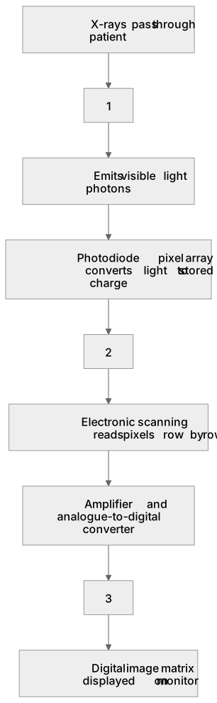
1Box 1
2Box 2
3Box 3
There is more to this lesson
The full lesson goes on to check your understanding and help you earn the marks. Create a free account to carry on, with your progress saved.
A week of Premium from your first lesson · Then the free plan continues · No card, no charge
Test your understanding of Image detection and enhancement (A-level only): Flat panel (FTP) detector including X-ray scintillator, photodiode pixels, electronic scanning.
Active recall beats re-reading. Take an instant quiz now to test what you’ve learned and lock it into long-term memory.
A week of Premium from your first lesson · Then the free plan continues · No card, no charge