AQA · A-Level · Physics
Course version: 7408 · Version from Sept 2016
Students will be required to make comparisons between imaging techniques.
Full specification wording
Imaging comparisons (A-level only): Students will be required to make comparisons between imaging techniques.
Comparing imaging techniques means judging each method against the clinical question, not simply listing its properties.
Checked against the AQA 7408 specification (opens in a new tab) on .
- Point 1 of 2
- 4 key terms
Study notes
From the checked teaching notes for this specification point.
The idea
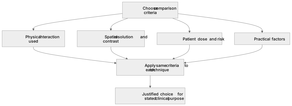
Comparing imaging techniques means judging each method against the clinical question, not simply listing its properties. A useful comparison selects criteria such as the physical interaction used, the type of radiation, spatial resolution, contrast mechanism, whether the image is structural or functional, the radiation dose to the patient, cost, availability, speed and patient experience. For example, X-ray imaging relies on differential attenuation and gives good bone detail with a moderate dose, whereas ultrasound uses reflected high-frequency sound, involves no ionising radiation and is well suited to soft tissue and real-time imaging, but cannot penetrate bone or gas-filled regions well. A strong answer applies the same criteria to each technique and reaches a justified conclusion about which is more suitable for a stated clinical situation.
What earns the marks
- Identify the physical principle of each technique, such as differential attenuation for X-rays, annihilation photon detection in PET, or reflection of ultrasound at tissue boundaries.
- Compare spatial resolution and contrast, explaining why one technique shows particular tissues or functional information better than another.
- Compare patient dose and risk, noting that techniques using ionising radiation deliver a dose whereas ultrasound and MRI do not.
- Compare practical factors such as cost, portability, availability, scan time and patient comfort or safety.
- Use consistent criteria across the techniques being compared so that the comparison is fair and leads to a justified choice.
- Relate the comparison to a specific clinical purpose, such as imaging a suspected fracture, a soft-tissue injury or metabolically active tissue.
Common mistakes and how to put them right
Listing features of each technique separately without comparing them.
Put it right by applying the same criteria to each technique in turn.
Claiming that MRI uses ionising radiation.
Put it right by stating that MRI uses strong magnetic fields and radio waves, not ionising radiation.
Treating higher resolution as automatically better in every case.
Put it right by linking the choice of technique to the clinical question and the information required.
Exam tips
- Choose three or four comparison criteria and use them consistently for every technique discussed.
- Include at least one quantitative or semi-quantitative point, such as relative dose or typical resolution, where you can do so accurately.
- Finish with a justified recommendation for a stated clinical scenario rather than a neutral list.
Try the lesson
The start of the Imaging comparisons (A-level only): Students will be required to make comparisons between imaging techniques. lesson, made from checked teaching notes for this specification point.
Have a go first
Which imaging technique uses ionising radiation to produce an image?
Listen
Comparing imaging techniques means judging each method against the clinical question, not simply listing its properties. A useful comparison selects criteria such as the physical interaction used, the type of radiation, spatial resolution, contrast mechanism, whether the image is structural or functional, the radiation dose to the patient, cost, availability, speed and patient experience. For example, X-ray imaging relies on differential attenuation and gives good bone detail with a moderate dose, whereas ultrasound uses reflected high-frequency sound, involves no ionising radiation and is well suited to soft tissue and real-time imaging, but cannot penetrate bone or gas-filled regions well. A strong answer applies the same criteria to each technique and reaches a justified conclusion about which is more suitable for a stated clinical situation. A strong answer earns marks for these points. Identify the physical principle of each technique, such as differential attenuation for X-rays, annihilation photon detection in PET, or reflection of ultrasound at tissue boundaries. Compare spatial resolution and contrast, explaining why one technique shows particular tissues or functional information better than another. Compare patient dose and risk, noting that techniques using ionising radiation deliver a dose whereas ultrasound and MRI do not. Compare practical factors such as cost, portability, availability, scan time and patient comfort or safety. A common slip: Listing features of each technique separately without comparing them. Instead, applying the same criteria to each technique in turn. Exam tip: Choose three or four comparison criteria and use them consistently for every technique discussed.
See it
Key words
Say what each word means before you turn the card.
Physical principles of medical imaging
Try answering aloud or in your head before turning the card.
Image quality and contrast mechanisms
Try answering aloud or in your head before turning the card.
Patient dose and radiation risk
Try answering aloud or in your head before turning the card.
Clinical suitability and practical constraints
Try answering aloud or in your head before turning the card.
See every definition
- Physical principles of medical imaging
- The physical interaction each technique uses, such as differential attenuation for X-rays, annihilation photon detection in PET, or reflection of ultrasound at tissue boundaries.
- Image quality and contrast mechanisms
- Spatial resolution and contrast, and why one technique shows particular tissues or functional information better than another.
- Patient dose and radiation risk
- Techniques using ionising radiation deliver a dose whereas ultrasound and MRI do not.
- Clinical suitability and practical constraints
- Practical factors such as cost, portability, availability, scan time and patient comfort or safety, related to a specific clinical purpose.
Label the diagram
Some boxes have lost their names. Pick the right name for each number.
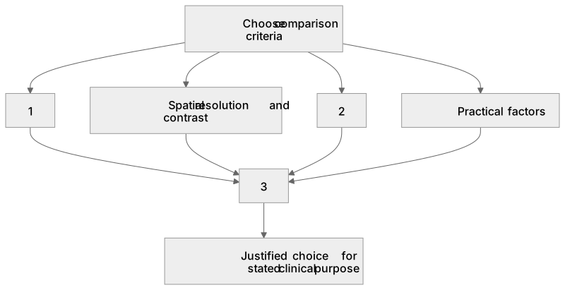
1Box 1
2Box 2
3Box 3
There is more to this lesson
The full lesson goes on to check your understanding and help you earn the marks. Create a free account to carry on, with your progress saved.
A week of Premium from your first lesson · Then the free plan continues · No card, no charge
Test your understanding of Imaging comparisons (A-level only): Students will be required to make comparisons between imaging techniques.
Active recall beats re-reading. Take an instant quiz now to test what you’ve learned and lock it into long-term memory.
A week of Premium from your first lesson · Then the free plan continues · No card, no charge