AQA · A-Level · Physics
Course version: 7408 · Version from Sept 2016
The properties should include the radiation emitted, the half-life, the…
Full specification wording
Imaging techniques (A-level only): The properties should include the radiation emitted, the half-life, the energy of the gamma radiation, the ability for it to be labelled with a compound with an affinity for a particular organ.
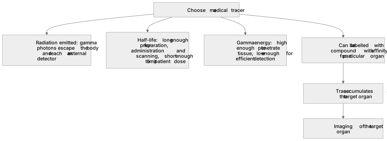
Choosing a medical tracer means matching four properties to the imaging task.
Checked against the AQA 7408 specification (opens in a new tab) on .
- Point 1 of 2
- 4 key terms
Study notes
From the checked teaching notes for this specification point.
The idea
Choosing a medical tracer means matching four properties to the imaging task. The emitted radiation must escape the body and be detected externally, so gamma photons are used rather than alpha or beta particles, which are absorbed before reaching a detector. The half-life must be long enough for preparation, administration and scanning, yet short enough to limit patient dose; hours are typical. Gamma energy must be high enough to penetrate tissue but low enough for efficient detection, so a value around 100 keV is suitable. Finally, the isotope must be attachable to a pharmaceutical with affinity for the target organ, so it concentrates where imaging is needed.
For example: Technetium-99m labelled to a phosphate compound accumulates in bone, allowing a bone scan.
What earns the marks
- Gamma emission is required because gamma photons penetrate tissue and reach an external detector, whereas alpha and beta radiation is largely absorbed within the body.
- The half-life must be a compromise: long enough to allow production, labelling, administration and imaging, but short enough that activity falls quickly and patient dose stays low.
- Gamma photon energy must be sufficient to penetrate the body yet suitable for efficient detection, so an intermediate energy such as about 100 keV is chosen.
- The isotope must be capable of being chemically bound to a compound that has an affinity for the organ of interest, so the tracer accumulates at the target site.
- A named example, such as technetium-99m attached to a bone-seeking phosphate compound, shows how the four properties combine in practice.
- The choice is assessed by explaining each property in relation to the imaging requirement, not by listing properties without justification.
Common mistakes and how to put them right
Choosing an alpha emitter because it is ionising.
Put it right: Select a gamma emitter.
Treating a very long half-life as automatically safer.
Put it right: Choose a half-life matched to the imaging procedure.
Assuming any isotope can be attached to any drug.
Put it right: Check that a suitable labelled compound can be formed.
Exam tips
- Link each property to its purpose in one sentence, for example penetration for detection or half-life for dose control.
- Use one worked example throughout, such as technetium-99m labelled to a bone-seeking compound, to show the properties working together.
- When comparing isotopes, state the imaging requirement first and then judge each isotope against it.
Try the lesson
The start of the Imaging techniques (A-level only): The properties should include the radiation emitted, the half-life, the energy of the gamma radiation, the ability for it to be labelled with a compound with an affinity for a particular organ. lesson, made from checked teaching notes for this specification point.
Have a go first
Which property makes technetium-99m suitable for use as a medical tracer in a gamma camera?
Listen
Choosing a medical tracer means matching four properties to the imaging task. The emitted radiation must escape the body and be detected externally, so gamma photons are used rather than alpha or beta particles, which are absorbed before reaching a detector. The half-life must be long enough for preparation, administration and scanning, yet short enough to limit patient dose; hours are typical. Gamma energy must be high enough to penetrate tissue but low enough for efficient detection, so a value around 100 keV is suitable. Finally, the isotope must be attachable to a pharmaceutical with affinity for the target organ, so it concentrates where imaging is needed. For example, technetium-99m labelled to a phosphate compound accumulates in bone, allowing a bone scan. A strong answer earns marks for these points. Gamma emission is required because gamma photons penetrate tissue and reach an external detector, whereas alpha and beta radiation is largely absorbed within the body. The half-life must be a compromise: long enough to allow production, labelling, administration and imaging, but short enough that activity falls quickly and patient dose stays low. Gamma photon energy must be sufficient to penetrate the body yet suitable for efficient detection, so an intermediate energy such as about 100 keV is chosen. The isotope must be capable of being chemically bound to a compound that has an affinity for the organ of interest, so the tracer accumulates at the target site. A common slip: Choosing an alpha emitter because it is ionising: alpha particles are stopped by tissue, so they cannot be detected outside the body. Instead, select a gamma emitter. Exam tip: Link each property to its purpose in one sentence, for example penetration for detection or half-life for dose control.
See it
Key words
Say what each word means before you turn the card.
Penetrating power and external detection
Try answering aloud or in your head before turning the card.
Balance of half-life against patient dose
Try answering aloud or in your head before turning the card.
Gamma photon energy and detector efficiency
Try answering aloud or in your head before turning the card.
Chemical labelling and organ specificity
Try answering aloud or in your head before turning the card.
See every definition
- Penetrating power and external detection
- Gamma photons penetrate tissue and reach an external detector, whereas alpha and beta radiation is largely absorbed within the body.
- Balance of half-life against patient dose
- The half-life must be long enough to allow production, labelling, administration and imaging, but short enough that activity falls quickly and patient dose stays low.
- Gamma photon energy and detector efficiency
- The energy must be sufficient to penetrate the body yet suitable for efficient detection, so an intermediate energy such as about 100 keV is chosen.
- Chemical labelling and organ specificity
- The isotope must be capable of being chemically bound to a compound that has an affinity for the organ of interest, so the tracer accumulates at the target site.
Label the diagram
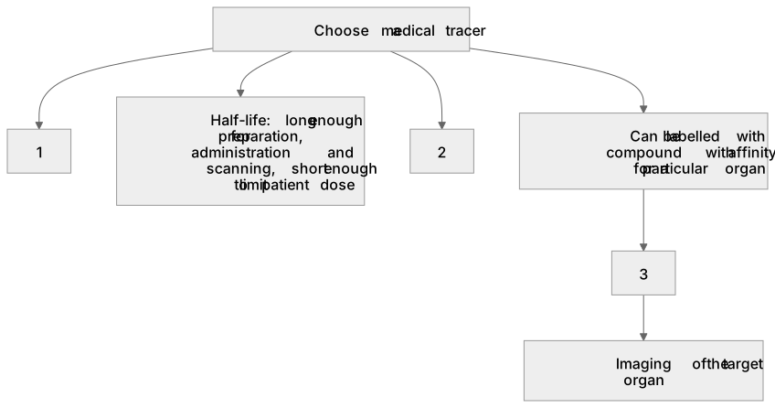
Some boxes have lost their names. Pick the right name for each number.
1Box 1
2Box 2
3Box 3
There is more to this lesson
The full lesson goes on to check your understanding and help you earn the marks. Create a free account to carry on, with your progress saved.
A week of Premium from your first lesson · Then the free plan continues · No card, no charge
Test your understanding of Imaging techniques (A-level only): The properties should include the radiation emitted, the half-life, the energy of the gamma radiation, the ability for it to be labelled with a compound with an affinity for a particular organ.
Active recall beats re-reading. Take an instant quiz now to test what you’ve learned and lock it into long-term memory.
A week of Premium from your first lesson · Then the free plan continues · No card, no charge