AQA · A-Level · Physics
Course version: 7408 · Version from Sept 2016
Ultrasound imaging (A-level only): Examples of applications.
Ultrasound imaging is used widely in medicine because it is non-ionising and can clearly differentiate soft tissue boundaries.
Checked against the AQA 7408 specification (opens in a new tab) on .
- Point 5 of 5
- 3 key terms
Study notes
From the checked teaching notes for this specification point.
The idea
Ultrasound imaging is used widely in medicine because it is non-ionising and can clearly differentiate soft tissue boundaries. Medical applications include prenatal scanning to monitor foetal growth and position, echocardiography to view heart valves and chambers in real time, and A-scans of the eye to measure axial length before lens implantation. In each case, a transducer emits high-frequency sound pulses into the body. Echoes return from boundaries where the acoustic impedance changes between tissues. The time delay of the echoes provides depth information, while the amplitude indicates the nature of the boundary. A coupling gel is used to match acoustic impedance and prevent reflection at the skin.
What earns the marks
- States at least two valid medical applications of ultrasound imaging, such as prenatal scanning, echocardiography, or eye measurement.
- Explains that ultrasound is used because it is non-ionising and therefore safer than X-rays for repeated or prenatal imaging.
- Links applications to the pulse–echo principle: a pulse is reflected at a boundary where acoustic impedance changes, and the echo time gives depth.
- Describes how a coupling medium such as gel is used to reduce reflection at the transducer–skin boundary and transmit ultrasound into the body.
- Explains how the amplitude of the returning echo depends on the difference in acoustic impedance between two media.
- Applies the depth equation distance = speed × time ÷ 2 to a stated medical application, for example measuring the distance to a foetal skull.
Common mistakes and how to put them right
Including industrial applications like testing welds.
Put it right by focusing exclusively on medical applications such as echocardiography for this option.
Claiming ultrasound is ionising radiation.
Put it right by stating ultrasound consists of longitudinal pressure waves and is non-ionising.
Using the one-way distance equation.
Put it right by calculating depth as speed × time ÷ 2, since the pulse travels to the boundary and back.
Exam tips
- When asked for applications, give named medical examples and state the physical principle each relies on, rather than listing names alone.
- Use the phrase non-ionising when explaining the safety advantage of ultrasound over X-rays.
Try the lesson
The start of the Ultrasound imaging (A-level only): Examples of applications. lesson, made from checked teaching notes for this specification point.
Have a go first
Which application commonly uses ultrasound imaging rather than X-ray imaging?
Listen
Ultrasound imaging is used widely in medicine because it is non-ionising and can clearly differentiate soft tissue boundaries. Medical applications include prenatal scanning to monitor foetal growth and position, echocardiography to view heart valves and chambers in real time, and A-scans of the eye to measure axial length before lens implantation. In each case, a transducer emits high-frequency sound pulses into the body. Echoes return from boundaries where the acoustic impedance changes between tissues. The time delay of the echoes provides depth information, while the amplitude indicates the nature of the boundary. A coupling gel is used to match acoustic impedance and prevent reflection at the skin. A strong answer earns marks for these points. States at least two valid medical applications of ultrasound imaging, such as prenatal scanning, echocardiography, or eye measurement. Explains that ultrasound is used because it is non-ionising and therefore safer than X-rays for repeated or prenatal imaging. Links applications to the pulse–echo principle: a pulse is reflected at a boundary where acoustic impedance changes, and the echo time gives depth. Describes how a coupling medium such as gel is used to reduce reflection at the transducer–skin boundary and transmit ultrasound into the body. A common slip: Including industrial applications like testing welds. Instead, focusing exclusively on medical applications such as echocardiography for this option. Exam tip: When asked for applications, give named medical examples and state the physical principle each relies on, rather than listing names alone.
See it
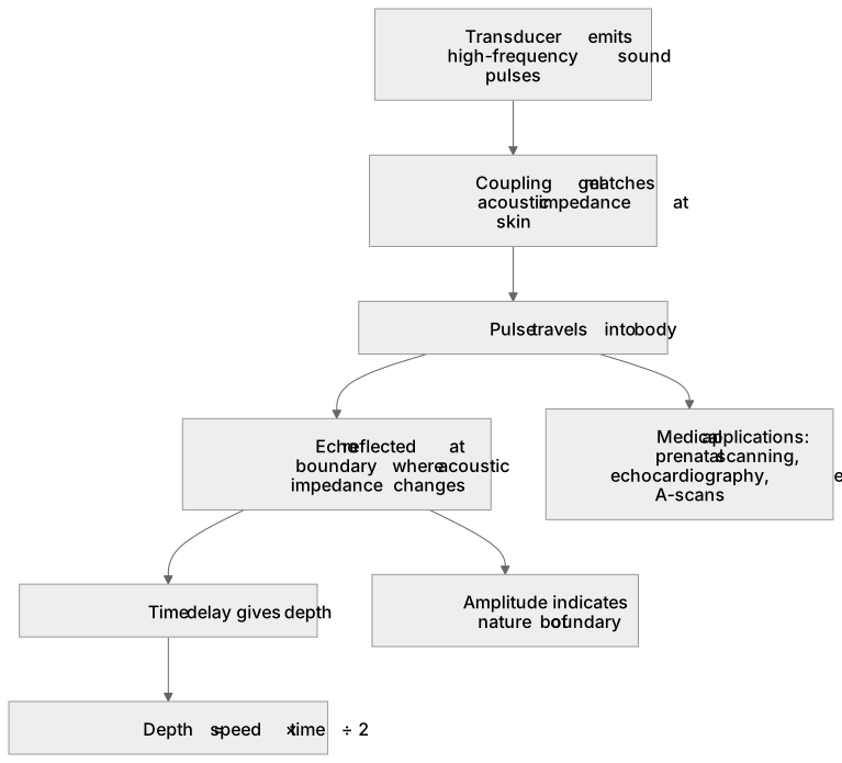
Key words
Say what each word means before you turn the card.
Acoustic impedance
Try answering aloud or in your head before turning the card.
Pulse–echo depth measurement
Try answering aloud or in your head before turning the card.
Non-ionising radiation safety
Try answering aloud or in your head before turning the card.
See every definition
- Acoustic impedance
- Echoes return from boundaries where this changes between tissues, and its difference between two media determines the amplitude of the returning echo.
- Pulse–echo depth measurement
- A transducer emits high-frequency sound pulses into the body, and the time delay of the echoes provides depth information.
- Non-ionising radiation safety
- Ultrasound is used because it is non-ionising and therefore safer than X-rays for repeated or prenatal imaging.
Label the diagram
Some boxes have lost their names. Pick the right name for each number.
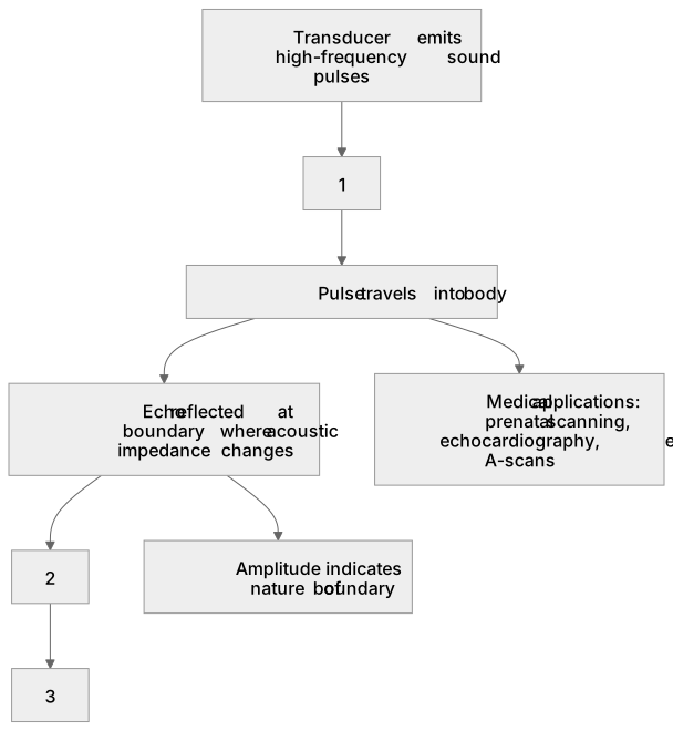
1Box 1
2Box 2
3Box 3
There is more to this lesson
The full lesson goes on to check your understanding and help you earn the marks. Create a free account to carry on, with your progress saved.
A week of Premium from your first lesson · Then the free plan continues · No card, no charge
Test your understanding of Ultrasound imaging (A-level only): Examples of applications.
Active recall beats re-reading. Take an instant quiz now to test what you’ve learned and lock it into long-term memory.
A week of Premium from your first lesson · Then the free plan continues · No card, no charge