AQA · A-Level · Physics
Course version: 7408 · Version from Sept 2016
Ultrasound imaging (A-level only): A-scans and B-scans.
An A-scan is a one-dimensional amplitude–time display: a pulse is emitted, and echoes from boundaries are shown as vertical spikes whose height gives echo amplitude and whose horizontal position…
Checked against the AQA 7408 specification (opens in a new tab) on .
- Point 4 of 5
- 4 key terms
Study notes
From the checked teaching notes for this specification point.
The idea
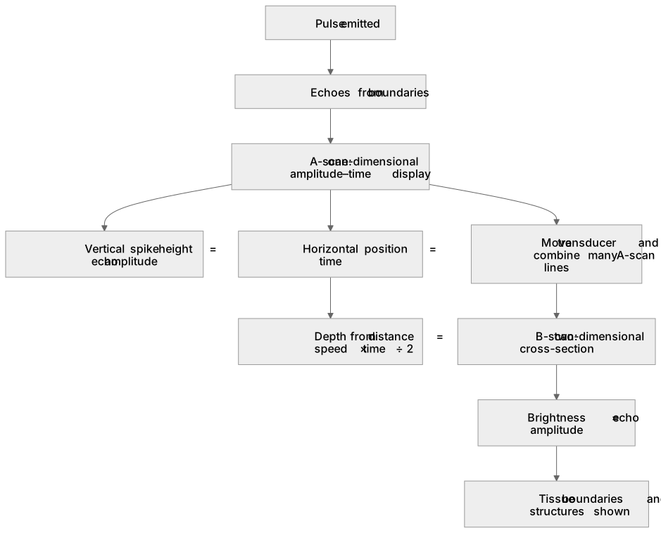
An A-scan is a one-dimensional amplitude–time display: a pulse is emitted, and echoes from boundaries are shown as vertical spikes whose height gives echo amplitude and whose horizontal position gives time, hence depth via distance = speed × time ÷ 2. A B-scan builds a two-dimensional cross-section by moving the transducer and combining many A-scan lines; echo brightness, not spike height, represents amplitude, so a B-scan image shows tissue boundaries and structures. For example, an A-scan can measure eye axial length from a corneal and retinal echo, while a B-scan can show a foetal head outline. Both use the same pulse–echo principle and the same speed of sound in tissue.
What earns the marks
- Describes an A-scan as a one-dimensional amplitude–time graph in which echo spikes are plotted against time.
- Explains that in an A-scan the vertical spike height represents echo amplitude and the horizontal position represents time, allowing depth to be calculated using distance = speed × time ÷ 2.
- Describes a B-scan as a two-dimensional image formed by moving the transducer and combining many A-scan lines.
- Explains that in a B-scan the brightness of each point represents echo amplitude, so boundaries between tissues of different acoustic impedance appear as bright lines.
- Applies the pulse–echo principle to both scans, including the need for a coupling medium and the use of the speed of sound in tissue.
- Compares the information provided: A-scans give precise depth measurements along one line, while B-scans give spatial structure across a plane.
Common mistakes and how to put them right
Confusing the axes of an A-scan.
Put it right: The horizontal axis is time, and depth is calculated from time using distance = speed × time ÷ 2.
Stating that a B-scan uses spike height to show echo amplitude.
Put it right: A B-scan uses brightness to represent echo amplitude.
Forgetting the factor of two in depth calculations.
Put it right: The pulse travels to the boundary and back, so distance = speed × time ÷ 2.
Exam tips
- When describing an A-scan, always state what the vertical and horizontal axes represent before interpreting a trace.
- When calculating depth from an echo time, write the full equation distance = speed × time ÷ 2 and substitute values with units.
- When comparing A-scans and B-scans, use the terms one-dimensional and two-dimensional explicitly and link each to its display method.
Try the lesson
The start of the Ultrasound imaging (A-level only): A-scans and B-scans. lesson, made from checked teaching notes for this specification point.
Have a go first
In ultrasound imaging, which type of scan displays the reflected pulses as a one-dimensional graph of signal amplitude against time?
Listen
An A-scan is a one-dimensional amplitude–time display: a pulse is emitted, and echoes from boundaries are shown as vertical spikes whose height gives echo amplitude and whose horizontal position gives time, hence depth via distance = speed × time ÷ 2. A B-scan builds a two-dimensional cross-section by moving the transducer and combining many A-scan lines; echo brightness, not spike height, represents amplitude, so a B-scan image shows tissue boundaries and structures. For example, an A-scan can measure eye axial length from a corneal and retinal echo, while a B-scan can show a foetal head outline. Both use the same pulse–echo principle and the same speed of sound in tissue. A strong answer earns marks for these points. Describes an A-scan as a one-dimensional amplitude–time graph in which echo spikes are plotted against time. Explains that in an A-scan the vertical spike height represents echo amplitude and the horizontal position represents time, allowing depth to be calculated using distance = speed × time ÷ 2. Describes a B-scan as a two-dimensional image formed by moving the transducer and combining many A-scan lines. Explains that in a B-scan the brightness of each point represents echo amplitude, so boundaries between tissues of different acoustic impedance appear as bright lines. A common slip: Confusing the axes of an A-scan: the error is treating the horizontal axis as depth directly. Instead, the horizontal axis is time, and depth is calculated from time using distance = speed × time ÷ 2. Exam tip: When describing an A-scan, always state what the vertical and horizontal axes represent before interpreting a trace.
See it
Key words
Say what each word means before you turn the card.
A-scan
Try answering aloud or in your head before turning the card.
B-scan
Try answering aloud or in your head before turning the card.
Pulse–echo principle
Try answering aloud or in your head before turning the card.
Depth calculation from echo time
Try answering aloud or in your head before turning the card.
See every definition
- A-scan
- A one-dimensional amplitude–time display in which a pulse is emitted and echoes from boundaries are shown as vertical spikes.
- B-scan
- A two-dimensional cross-section built by moving the transducer and combining many A-scan lines, with echo brightness representing amplitude.
- Pulse–echo principle
- A pulse is emitted and echoes from boundaries are detected, used by both A-scans and B-scans with the same speed of sound in tissue.
- Depth calculation from echo time
- Depth is found from the echo time using distance = speed × time ÷ 2, since the pulse travels to the boundary and back.
Label the diagram
Some boxes have lost their names. Pick the right name for each number.
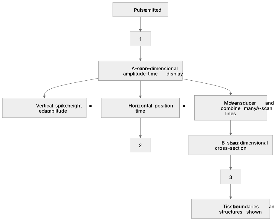
1Box 1
2Box 2
3Box 3
There is more to this lesson
The full lesson goes on to check your understanding and help you earn the marks. Create a free account to carry on, with your progress saved.
A week of Premium from your first lesson · Then the free plan continues · No card, no charge
Test your understanding of Ultrasound imaging (A-level only): A-scans and B-scans.
Active recall beats re-reading. Take an instant quiz now to test what you’ve learned and lock it into long-term memory.
A week of Premium from your first lesson · Then the free plan continues · No card, no charge