AQA · GCSE · Physics
Course version: 8463 · Version from Sept 2016
Nuclear radiations are used in medicine for the: exploration of internal…
Full specification wording
Uses of nuclear radiation: Nuclear radiations are used in medicine for the: exploration of internal organs control or destruction of unwanted tissue.
Medicine uses nuclear radiation in two distinct ways.
Checked against the AQA 8463 specification (opens in a new tab) on .
- Point 1 of 2
- 3 key terms
Study notes
From the checked teaching notes for this specification point.
The idea
Medicine uses nuclear radiation in two distinct ways. For exploration, a tracer containing a gamma-emitting isotope is swallowed or injected and taken up by the organ under investigation; because gamma radiation is weakly ionising and highly penetrating, it passes out of the body to a detector such as a gamma camera, building an image of how the organ is working with a modest dose to the patient. For control or destruction of unwanted tissue, high-energy gamma beams are aimed at a tumour from several directions so the beams cross only at the tumour, or a sealed source is implanted next to it. Alpha emitters are never used as tracers, since the radiation would be absorbed by tissue and never escape to be detected.
What earns the marks
- Describing a tracer as a gamma-emitting isotope taken into the body and followed by a detector outside it.
- Gamma being chosen because it penetrates out of the body and is weakly ionising.
- External radiotherapy directing gamma beams at a tumour, rotating or crossing them so healthy tissue receives less.
- Internal treatment using an implanted or injected source placed in or next to the unwanted tissue.
Common mistakes and how to put them right
Choosing an alpha emitter for a tracer, when the radiation could not leave the body.
Saying radiotherapy destroys only cancer cells and leaves healthy tissue untouched.
Describing a tracer as a treatment that cures the illness.
Confusing an X-ray image, which uses an external machine, with a gamma tracer scan.
Using a long half-life isotope as a tracer and so leaving the patient with a prolonged dose.
Exam tips
- Say what the radiation does, not just what the technique is called, because the mark is usually for the mechanism.
- Name the radiation type and give a reason for it; 'gamma, because it penetrates out of the body' is a complete point.
- Separate exploration from destruction in your answer, since the question usually wants one of each.
Try the lesson
The start of the Uses of nuclear radiation: Nuclear radiations are used in medicine for the: exploration of internal organs control or destruction of unwanted tissue. lesson, made from checked teaching notes for this specification point.
Have a go first
What are the two main uses of nuclear radiation in medicine?
Listen
Medicine uses nuclear radiation in two distinct ways. For exploration, a tracer containing a gamma-emitting isotope is swallowed or injected and taken up by the organ under investigation; because gamma radiation is weakly ionising and highly penetrating, it passes out of the body to a detector such as a gamma camera, building an image of how the organ is working with a modest dose to the patient. For control or destruction of unwanted tissue, high-energy gamma beams are aimed at a tumour from several directions so the beams cross only at the tumour, or a sealed source is implanted next to it. Alpha emitters are never used as tracers, since the radiation would be absorbed by tissue and never escape to be detected. A strong answer earns marks for these points. describing a tracer as a gamma-emitting isotope taken into the body and followed by a detector outside it. gamma being chosen because it penetrates out of the body and is weakly ionising. external radiotherapy directing gamma beams at a tumour, rotating or crossing them so healthy tissue receives less. internal treatment using an implanted or injected source placed in or next to the unwanted tissue. Exam tip: Say what the radiation does, not just what the technique is called, because the mark is usually for the mechanism.
See it
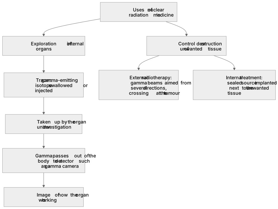
Key words
Say what each word means before you turn the card.
medical tracers
Try answering aloud or in your head before turning the card.
gamma cameras
Try answering aloud or in your head before turning the card.
radiotherapy
Try answering aloud or in your head before turning the card.
See every definition
- medical tracers
- A gamma-emitting isotope is taken into the body and followed by a detector outside it, building an image of how an organ is working.
- gamma cameras
- A detector outside the body that receives gamma radiation passing out of the body and builds an image of how the organ is working.
- radiotherapy
- High-energy gamma beams are aimed at a tumour from several directions so the beams cross only at the tumour, or a sealed source is implanted next to it.
Label the diagram
Some boxes have lost their names. Pick the right name for each number.
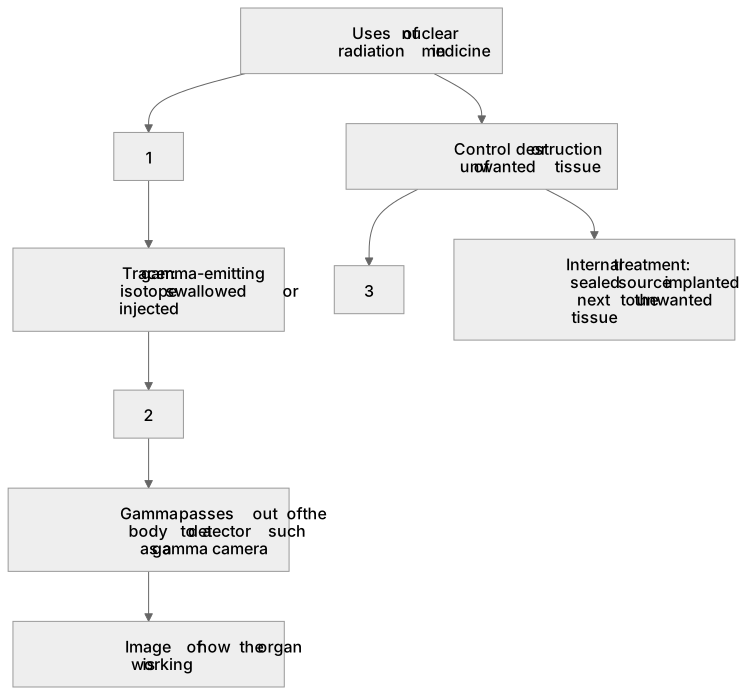
1Box 1
2Box 2
3Box 3
There is more to this lesson
The full lesson goes on to check your understanding and help you earn the marks. Create a free account to carry on, with your progress saved.
A week of Premium from your first lesson · Then the free plan continues · No card, no charge
Test your understanding of Uses of nuclear radiation: Nuclear radiations are used in medicine for the: exploration of internal organs control or destruction of unwanted tissue.
Active recall beats re-reading. Take an instant quiz now to test what you’ve learned and lock it into long-term memory.
A week of Premium from your first lesson · Then the free plan continues · No card, no charge