AQA · A-Level · Biology
Course version: 7402 · Version from Sept 2016
The response of T lymphocytes to a foreign antigen (the cellular response).
Full specification wording
Cell recognition and the immune system: The response of T lymphocytes to a foreign antigen (the cellular response).
T lymphocytes mature in the thymus, each carrying receptors of a single specific tertiary structure.
Checked against the AQA 7402 specification (opens in a new tab) on .
- Point 4 of 18
- 4 key terms
Study notes
From the checked teaching notes for this specification point.
The idea
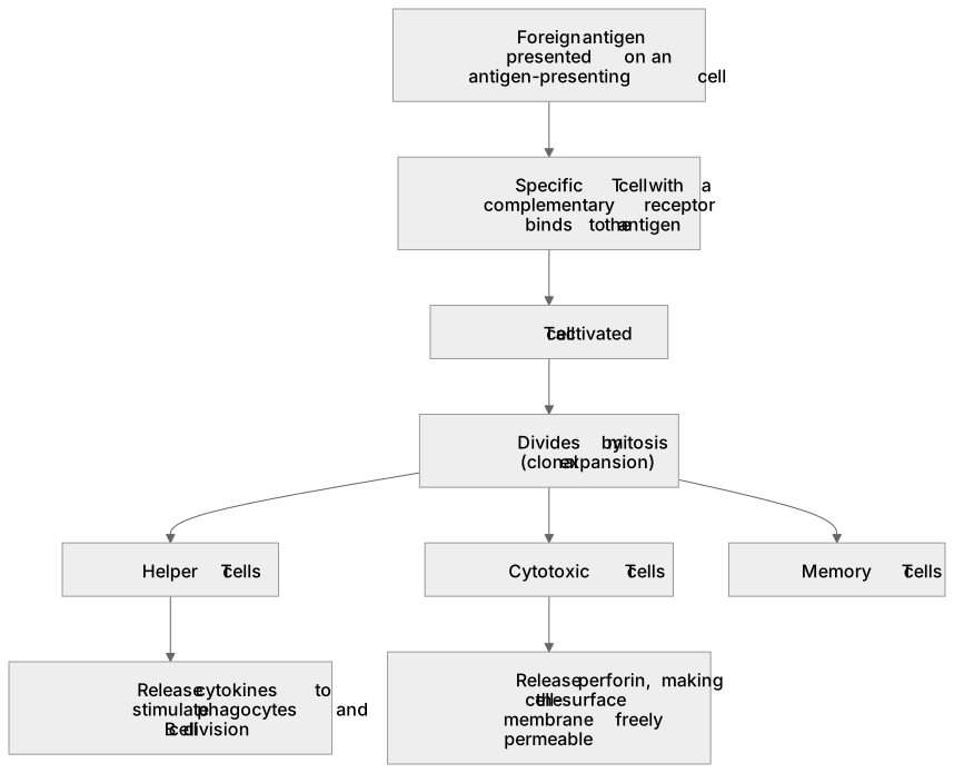
T lymphocytes mature in the thymus, each carrying receptors of a single specific tertiary structure. They respond only to antigens presented on a cell surface, targeting virus-infected cells or transplanted tissue. When an antigen-presenting cell displays a foreign antigen, a T cell with a complementary receptor binds to it. This activates the T cell, causing it to divide by mitosis (clonal expansion). The resulting clone is lineage-specific: activated helper T cells release cytokines to stimulate phagocytes and B cell division, while cytotoxic T cells kill infected cells by releasing perforin, making the cell-surface membrane freely permeable. Both lineages also produce memory T cells for a rapid secondary response.
What earns the marks
- A foreign or faulty protein being recognised as an antigen on an antigen-presenting cell.
- A specific T cell with a complementary receptor binding to the presented antigen.
- The activated T cell dividing by mitosis to form a clone (clonal expansion).
- Helper T cells releasing cytokines that stimulate phagocytes and B cell division.
- Cytotoxic T cells releasing perforin to make the infected cell's membrane freely permeable.
Common mistakes and how to put them right
Stating that T cells produce antibodies.
Put it right: Only plasma cells derived from B cells secrete antibodies.
Claiming that helper T cells stimulate clonal selection of B cells.
Put it right: Antigens cause selection, while helper T cells stimulate clonal expansion.
Stating that cytotoxic T cells 'kill the cell' without a mechanism.
Put it right: You must specify that perforin makes the membrane freely permeable.
Exam tips
- Always use 'complementary' when describing the fit between the T cell receptor and the antigen; 'same shape' will not be credited.
- When asked how the immune system destroys abnormal cells, start by stating that the abnormal protein is recognised as a foreign antigen.
Try the lesson
The start of the Cell recognition and the immune system: The response of T lymphocytes to a foreign antigen (the cellular response). lesson, made from checked teaching notes for this specification point.
Have a go first
How does a helper T lymphocyte initially recognise a foreign antigen presented by a phagocyte?
Listen
T lymphocytes mature in the thymus, each carrying receptors of a single specific tertiary structure. They respond only to antigens presented on a cell surface, targeting virus-infected cells or transplanted tissue. When an antigen-presenting cell displays a foreign antigen, a T cell with a complementary receptor binds to it. This activates the T cell, causing it to divide by mitosis (clonal expansion). The resulting clone is lineage-specific: activated helper T cells release cytokines to stimulate phagocytes and B cell division, while cytotoxic T cells kill infected cells by releasing perforin, making the cell-surface membrane freely permeable. Both lineages also produce memory T cells for a rapid secondary response. A strong answer earns marks for these points. a foreign or faulty protein being recognised as an antigen on an antigen-presenting cell. a specific T cell with a complementary receptor binding to the presented antigen. the activated T cell dividing by mitosis to form a clone (clonal expansion). helper T cells releasing cytokines that stimulate phagocytes and B cell division. Exam tip: Always use 'complementary' when describing the fit between the T cell receptor and the antigen; 'same shape' will not be credited.
See it
Key words
Say what each word means before you turn the card.
clonal expansion
Try answering aloud or in your head before turning the card.
cytotoxic T cells
Try answering aloud or in your head before turning the card.
perforin
Try answering aloud or in your head before turning the card.
helper T cells
Try answering aloud or in your head before turning the card.
See every definition
- clonal expansion
- The activated T cell divides by mitosis to form a clone.
- cytotoxic T cells
- Cells that kill infected cells by releasing perforin, making the cell-surface membrane freely permeable.
- perforin
- Released by cytotoxic T cells to make the infected cell's membrane freely permeable.
- helper T cells
- Activated cells that release cytokines to stimulate phagocytes and B cell division.
Label the diagram
Some boxes have lost their names. Pick the right name for each number.
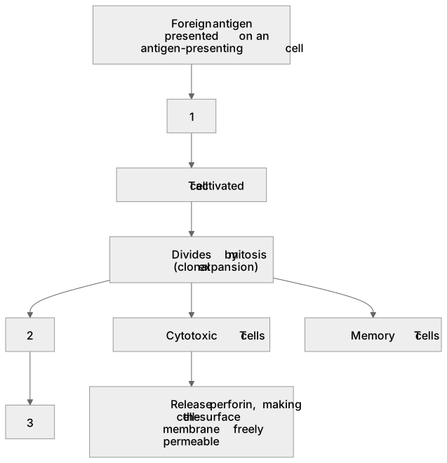
1Box 1
2Box 2
3Box 3
There is more to this lesson
The full lesson goes on to check your understanding and help you earn the marks. Create a free account to carry on, with your progress saved.
A week of Premium from your first lesson · Then the free plan continues · No card, no charge
Test your understanding of Cell recognition and the immune system: The response of T lymphocytes to a foreign antigen (the cellular response).
Active recall beats re-reading. Take an instant quiz now to test what you’ve learned and lock it into long-term memory.
A week of Premium from your first lesson · Then the free plan continues · No card, no charge