AQA · A-Level · Biology
Course version: 7402 · Version from Sept 2016
The role of antigen-presenting cells in the cellular response.
Full specification wording
Cell recognition and the immune system: The role of antigen-presenting cells in the cellular response.
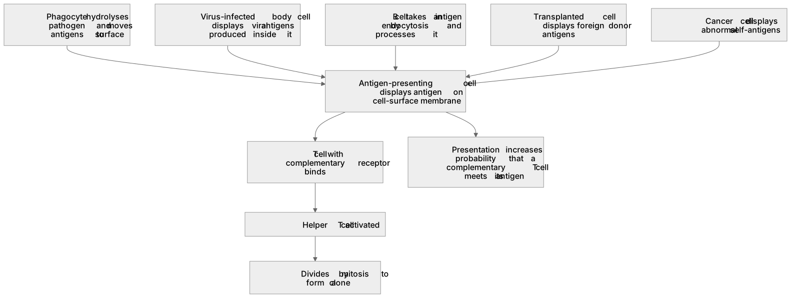
An antigen-presenting cell displays antigen on its cell-surface membrane, where a T cell with a complementary receptor can bind it.
Checked against the AQA 7402 specification (opens in a new tab) on .
- Point 5 of 18
- 4 key terms
Study notes
From the checked teaching notes for this specification point.
The idea
An antigen-presenting cell displays antigen on its cell-surface membrane, where a T cell with a complementary receptor can bind it. Sources include phagocytes that have hydrolysed a pathogen and moved its antigens to the surface; body cells infected by a virus, which display viral antigens produced inside them; and B cells that have taken in antigen by endocytosis and processed it. Transplanted cells display foreign donor antigens, while cancer cells display abnormal self-antigens. Presentation increases the probability that a complementary T cell meets its antigen: each T cell recognises only one antigen, cells carrying that receptor are rare, and free antigen in lymph is easily missed. Display on a cell surface in lymph nodes, through which lymphocytes circulate, raises the chance of contact. After binding, the helper T cell is activated and divides by mitosis to form a clone.
What earns the marks
- Define an antigen-presenting cell as a cell displaying antigen on its cell-surface membrane, and note that the displayed antigen may be foreign or an abnormal self-antigen.
- Name at least two sources and state how each acquires its antigen: a phagocyte after hydrolysing a pathogen, a virus-infected body cell, or a B cell that has taken in antigen by endocytosis.
- State that a T cell with a complementary receptor binds to the presented antigen.
- Explain that presentation increases the chance of a lymphocyte meeting its specific antigen, because each T cell recognises only one antigen and such cells are rare.
- State that the activated helper T cell divides by mitosis to form a clone.
- Distinguish transplanted cells, which present foreign donor antigens, from cancer cells, which present abnormal self-antigens.
Common mistakes and how to put them right
Assuming only phagocytes present antigens, and so losing the marks for infected body cells and B cells.
Put it right: Name at least two sources and state how each acquires its antigen.
Saying the antigen-presenting cell 'gives' the antigen to the T cell rather than displaying it for binding.
Put it right: The antigen is displayed on the membrane and the T cell's receptor binds to it.
Confusing antigen presentation with antibody production.
Put it right: Presentation is display of antigen; antibody production is secretion by plasma cells.
Describing antigen-presenting cells as a type of lymphocyte.
Put it right: Macrophages and infected body cells are not lymphocytes, though B cells are.
Describing the display of antigen but never stating the advantage it brings, which is where the explain mark sits.
Put it right: Finish with the increased probability of a complementary T cell meeting the antigen.
Stating that transplanted cells display their own abnormal antigens.
Put it right: Transplanted cells carry foreign donor antigens, whereas cancer cells display abnormal self-antigens.
Exam tips
- Finish an 'explain the role' answer with the consequence, activation of the T cell and clonal expansion, not just with the display of antigen.
- When asked to give cell types that stimulate an immune response, antigen-presenting cells such as macrophages and infected body cells are valid examples; check the wording of the question before committing.
Try the lesson
The start of the Cell recognition and the immune system: The role of antigen-presenting cells in the cellular response. lesson, made from checked teaching notes for this specification point.
Have a go first
Which cell presents antigens to T lymphocytes during the cellular response?
Listen
An antigen-presenting cell displays antigen on its cell-surface membrane, where a T cell with a complementary receptor can bind it. Sources include phagocytes that have hydrolysed a pathogen and moved its antigens to the surface; body cells infected by a virus, which display viral antigens produced inside them; and B cells that have taken in antigen by endocytosis and processed it. Transplanted cells display foreign donor antigens, while cancer cells display abnormal self-antigens. Presentation increases the probability that a complementary T cell meets its antigen: each T cell recognises only one antigen, cells carrying that receptor are rare, and free antigen in lymph is easily missed. Display on a cell surface in lymph nodes, through which lymphocytes circulate, raises the chance of contact. After binding, the helper T cell is activated and divides by mitosis to form a clone. A strong answer earns marks for these points. Define an antigen-presenting cell as a cell displaying antigen on its cell-surface membrane, and note that the displayed antigen may be foreign or an abnormal self-antigen. Name at least two sources and state how each acquires its antigen: a phagocyte after hydrolysing a pathogen, a virus-infected body cell, or a B cell that has taken in antigen by endocytosis. State that a T cell with a complementary receptor binds to the presented antigen. Explain that presentation increases the chance of a lymphocyte meeting its specific antigen, because each T cell recognises only one antigen and such cells are rare. A common slip: Assuming only phagocytes present antigens, and so losing the marks for infected body cells and B cells. Instead, name at least two sources and state how each acquires its antigen. Exam tip: Finish an 'explain the role' answer with the consequence, activation of the T cell and clonal expansion, not just with the display of antigen.
See it
Key words
Say what each word means before you turn the card.
antigen-presenting cell
Try answering aloud or in your head before turning the card.
virus-infected body cell
Try answering aloud or in your head before turning the card.
probability of a lymphocyte meeting its antigen
Try answering aloud or in your head before turning the card.
activation of helper T cells
Try answering aloud or in your head before turning the card.
See every definition
- antigen-presenting cell
- A cell displaying antigen on its cell-surface membrane, where the displayed antigen may be foreign or an abnormal self-antigen.
- virus-infected body cell
- A source of presented antigen that displays viral antigens produced inside it.
- probability of a lymphocyte meeting its antigen
- Increased by presentation, because each T cell recognises only one antigen, cells carrying that receptor are rare, and free antigen in lymph is easily missed.
- activation of helper T cells
- After binding to a presented antigen, the helper T cell is activated and divides by mitosis to form a clone.
Label the diagram
Some boxes have lost their names. Pick the right name for each number.
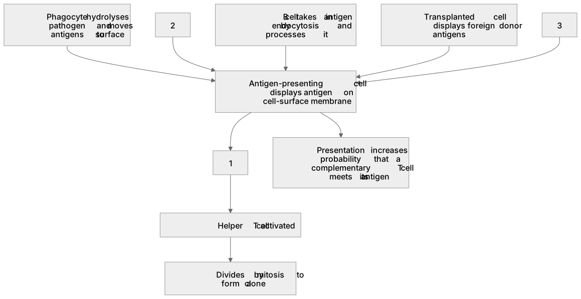
1Box 1
2Box 2
3Box 3
There is more to this lesson
The full lesson goes on to check your understanding and help you earn the marks. Create a free account to carry on, with your progress saved.
A week of Premium from your first lesson · Then the free plan continues · No card, no charge
Test your understanding of Cell recognition and the immune system: The role of antigen-presenting cells in the cellular response.
Active recall beats re-reading. Take an instant quiz now to test what you’ve learned and lock it into long-term memory.
A week of Premium from your first lesson · Then the free plan continues · No card, no charge