AQA · A-Level · Biology
Course version: 7402 · Version from Sept 2016
Mass transport in animals: The gross structure of the human heart.
The heart has four chambers: two thin-walled atria receiving blood and two thicker-walled ventricles pumping it out.
Checked against the AQA 7402 specification (opens in a new tab) on .
- Point 9 of 14
- 4 key terms
Study notes
From the checked teaching notes for this specification point.
The idea
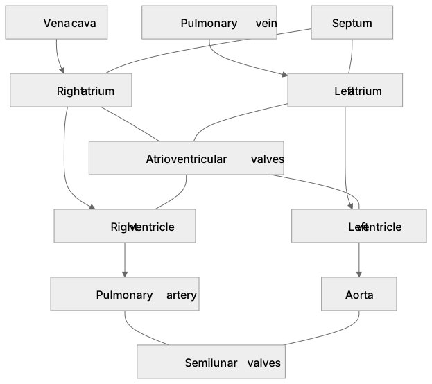
The heart has four chambers: two thin-walled atria receiving blood and two thicker-walled ventricles pumping it out. The vena cava brings deoxygenated blood to the right atrium; the pulmonary vein brings oxygenated blood to the left atrium. A septum separates the sides, preventing mixing. Atrioventricular valves separate atria and ventricles, anchored by tendinous cords that prevent inversion during ventricular contraction. Semilunar valves sit at the base of the pulmonary artery and aorta, preventing backflow into the ventricles. The left ventricle wall is much thicker than the right because it generates higher pressure to force blood around the systemic circulation. The right ventricle pumps at lower pressure, which protects the delicate pulmonary capillaries. Coronary arteries supply the cardiac muscle with oxygenated blood.
What earns the marks
- Identifies the vena cava and pulmonary vein as the major vessels returning blood to the right and left atria respectively.
- Explains that the left ventricle has a thicker muscular wall than the right ventricle to generate higher pressure for systemic circulation.
- States that the septum prevents oxygenated and deoxygenated blood from mixing.
- Describes the position of atrioventricular valves (between atria and ventricles) and semilunar valves (at the base of the aorta and pulmonary artery), and states their function in preventing backflow.
- Identifies coronary arteries as the vessels supplying oxygenated blood to cardiac muscle tissue.
Common mistakes and how to put them right
Labelling the heart from the reader's viewpoint.
Put it right: Label left and right from the heart's perspective, so the diagram's right is the heart's left.
Stating the left ventricle is larger because it holds more blood.
Put it right: It has a thicker muscular wall to generate higher pressure, but holds approximately the same volume as the right ventricle.
Confusing the major vessels.
Put it right: The vena cava and pulmonary vein bring blood into the atria, while the aorta and pulmonary artery take blood out of the ventricles.
Stating that lower right ventricular pressure ensures blood flows slowly enough for gas exchange.
Put it right: Lower pulmonary pressure mainly protects delicate capillaries; gas exchange depends on surface area, a thin diffusion barrier and partial-pressure gradients.
Exam tips
- When explaining ventricle wall thickness, always explicitly use the word 'pressure' rather than just saying it pumps blood 'further'.
- Note that the right ventricle pumps at a lower pressure to protect the delicate pulmonary capillaries, not to slow blood flow for gas exchange.
Try the lesson
The start of the Mass transport in animals: The gross structure of the human heart. lesson, made from checked teaching notes for this specification point.
Have a go first
Which blood vessel carries oxygenated blood away from the left ventricle of the human heart?
Listen
The heart has four chambers: two thin-walled atria receiving blood and two thicker-walled ventricles pumping it out. The vena cava brings deoxygenated blood to the right atrium; the pulmonary vein brings oxygenated blood to the left atrium. A septum separates the sides, preventing mixing. Atrioventricular valves separate atria and ventricles, anchored by tendinous cords that prevent inversion during ventricular contraction. Semilunar valves sit at the base of the pulmonary artery and aorta, preventing backflow into the ventricles. The left ventricle wall is much thicker than the right because it generates higher pressure to force blood around the systemic circulation. The right ventricle pumps at lower pressure, which protects the delicate pulmonary capillaries. Coronary arteries supply the cardiac muscle with oxygenated blood. A strong answer earns marks for these points. Identifies the vena cava and pulmonary vein as the major vessels returning blood to the right and left atria respectively. Explains that the left ventricle has a thicker muscular wall than the right ventricle to generate higher pressure for systemic circulation. States that the septum prevents oxygenated and deoxygenated blood from mixing. Describes the position of atrioventricular valves (between atria and ventricles) and semilunar valves (at the base of the aorta and pulmonary artery), and states their function in preventing backflow. A common slip: Labelling the heart from the reader's viewpoint. Instead, label left and right from the heart's perspective, so the diagram's right is the heart's left. Exam tip: When explaining ventricle wall thickness, always explicitly use the word 'pressure' rather than just saying it pumps blood 'further'.
See it
Key words
Say what each word means before you turn the card.
Atrioventricular valves
Try answering aloud or in your head before turning the card.
Semilunar valves
Try answering aloud or in your head before turning the card.
Septum
Try answering aloud or in your head before turning the card.
Coronary arteries
Try answering aloud or in your head before turning the card.
See every definition
- Atrioventricular valves
- These separate atria and ventricles and are anchored by tendinous cords that prevent inversion during ventricular contraction.
- Semilunar valves
- These sit at the base of the pulmonary artery and aorta, preventing backflow into the ventricles.
- Septum
- This separates the sides of the heart, preventing oxygenated and deoxygenated blood from mixing.
- Coronary arteries
- These supply the cardiac muscle with oxygenated blood.
Label the diagram
Some boxes have lost their names. Pick the right name for each number.
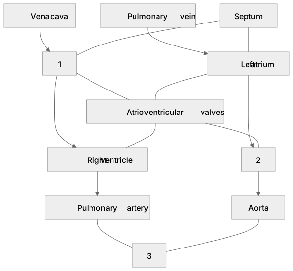
1Box 1
2Box 2
3Box 3
There is more to this lesson
The full lesson goes on to check your understanding and help you earn the marks. Create a free account to carry on, with your progress saved.
A week of Premium from your first lesson · Then the free plan continues · No card, no charge
Test your understanding of Mass transport in animals: The gross structure of the human heart.
Active recall beats re-reading. Take an instant quiz now to test what you’ve learned and lock it into long-term memory.
A week of Premium from your first lesson · Then the free plan continues · No card, no charge