Topic 8 – Exchange and transport in animals Revision Notes
Comprehensive revision notes for Edexcel GCSE.
Summary & Overview
Master the systems that keep you alive! This topic explores how animals exchange gases and transport nutrients around the body. It is heavily tested in exams, especially on linking structure to function in the heart, blood vessels, and lungs.
Study Material
## Overview
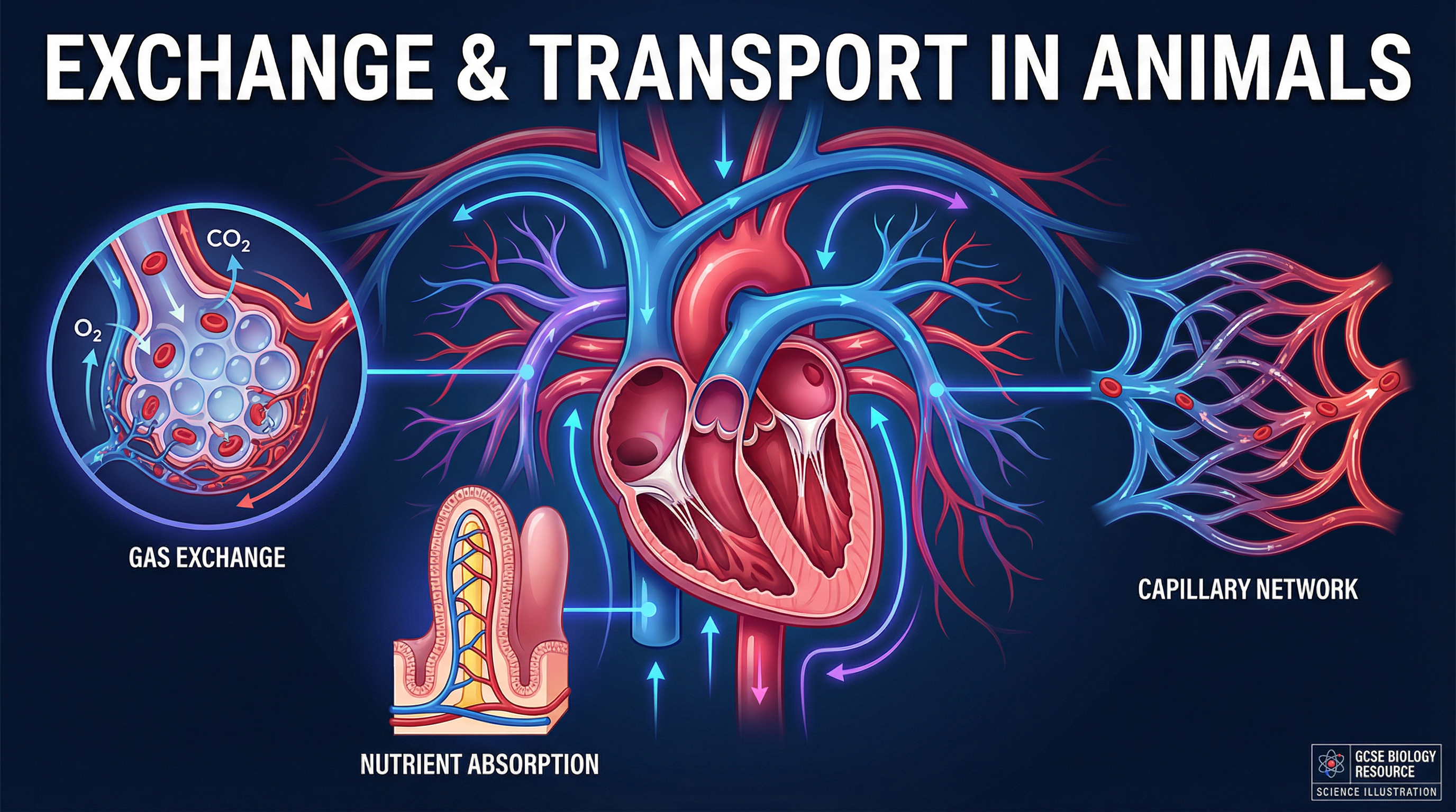
Welcome to Topic 8: Exchange and Transport in Animals. This crucial area of Biology explores how multicellular organisms solve the problem of size. While single-celled organisms can rely on simple diffusion to obtain oxygen and nutrients, large animals require specialised exchange surfaces and a mass transport system to survive.
Understanding this topic is essential for your exam success, as examiners frequently use it to test your ability to link structure to function (Assessment Objective 2). It connects synoptically to respiration (Topic 4) and non-communicable diseases like cardiovascular disease (Topic 7). Typical exam questions include explaining adaptations of alveoli, comparing blood vessels, and tracing the pathway of blood through the heart.
---
## Podcast Episode
Listen to our 10-minute deep dive into Exchange and Transport in Animals:

---
## Key Concepts
### Concept 1: The Need for Exchange Surfaces
As organisms get larger, their **surface area to volume ratio (SA:V)** decreases. This means the surface area available for diffusion is no longer sufficient to meet the metabolic demands of the large volume of cells inside the body. To overcome this, large animals have evolved specialised exchange surfaces, such as the lungs in humans.
**Example**: A single-celled bacterium has a large SA:V ratio, so oxygen can diffuse directly into the centre of the cell quickly enough for respiration. A human has a very small SA:V ratio, requiring lungs to provide a massive internal surface area for gas exchange.
### Concept 2: Gas Exchange at the Alveoli
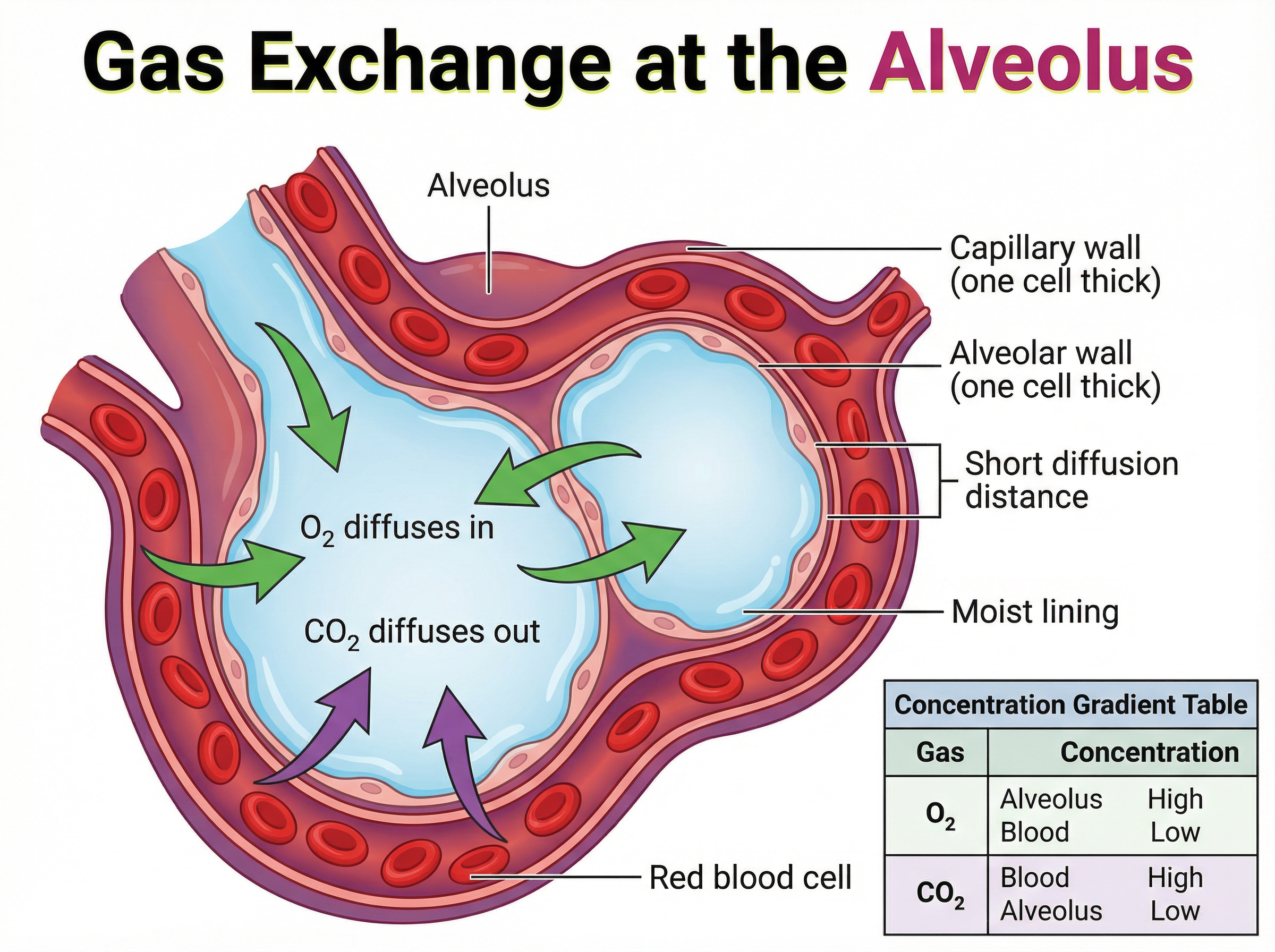
The lungs contain millions of tiny air sacs called **alveoli**, where gas exchange occurs. Oxygen diffuses from the air in the alveoli into the blood, while carbon dioxide diffuses from the blood into the alveoli to be exhaled.
The alveoli are perfectly adapted to maximise the rate of diffusion. They provide a massive surface area (roughly 70m² in humans). Their walls are just one cell thick, creating a very short diffusion pathway. They have a rich blood supply from a dense network of capillaries, which constantly removes oxygenated blood and brings in deoxygenated blood, maintaining a steep concentration gradient. Finally, they have a moist lining, allowing gases to dissolve before diffusing.
### Concept 3: The Double Circulatory System
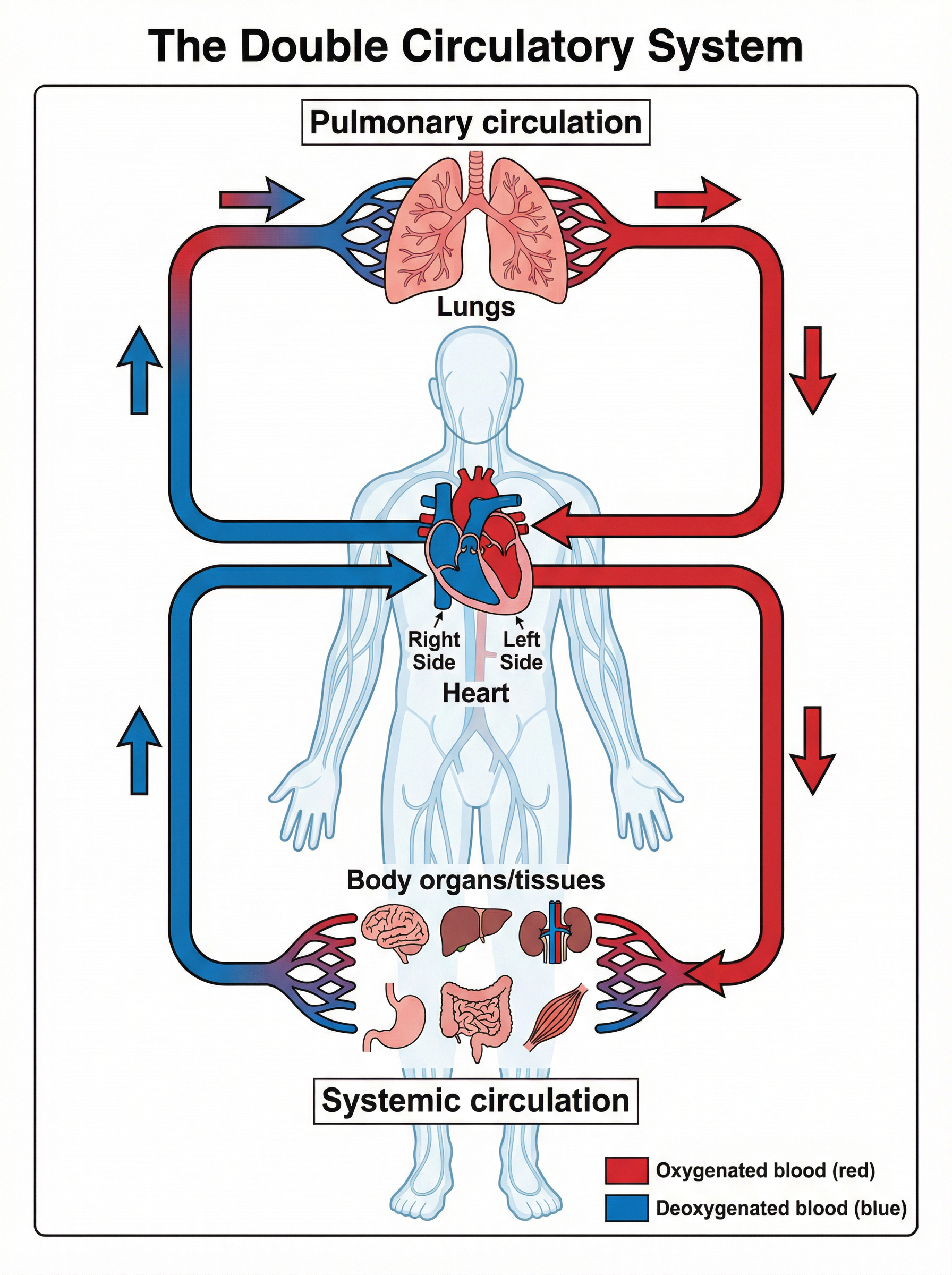
Humans have a **double circulatory system**, meaning blood passes through the heart twice for every complete circuit of the body.
1. **Pulmonary Circulation**: The right ventricle pumps deoxygenated blood to the lungs for gas exchange.
2. **Systemic Circulation**: The left ventricle pumps oxygenated blood to the rest of the body.
This system is highly efficient because blood returning from the lungs can be repressurised by the heart before being sent to the body, ensuring a fast delivery of oxygen to respiring tissues.
### Concept 4: Structure and Function of the Heart
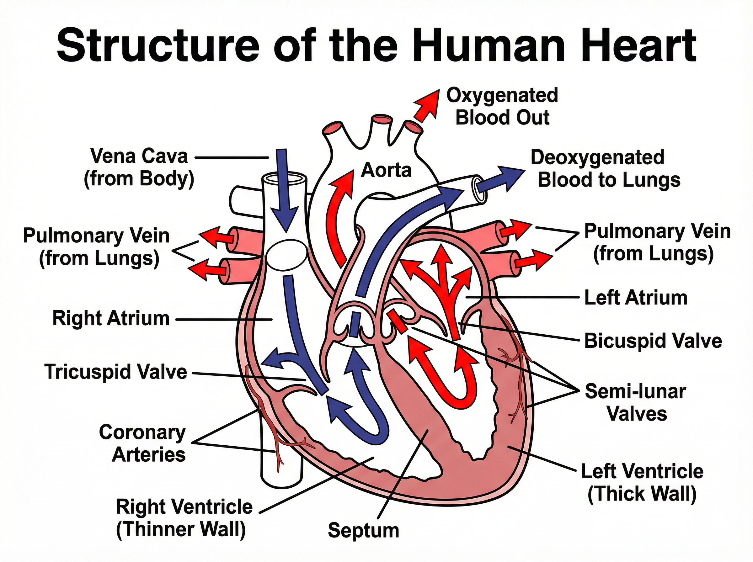
The heart is a muscular organ that pumps blood. It consists of four chambers: the right and left atria (top), and the right and left ventricles (bottom).
Deoxygenated blood enters the right atrium via the **vena cava**. It is pumped into the right ventricle, and then out to the lungs via the **pulmonary artery**. Oxygenated blood returns to the left atrium via the **pulmonary vein**. It is pumped into the left ventricle, and then out to the body via the **aorta**.
The **left ventricle** has a much thicker muscular wall than the right ventricle because it must generate a higher pressure to pump blood around the entire body, whereas the right ventricle only pumps blood to the nearby lungs. Valves (tricuspid, bicuspid, and semi-lunar) ensure blood flows in one direction and prevent backflow.
### Concept 5: Blood Vessels
There are three main types of blood vessels, each adapted to its function:
* **Arteries**: Carry blood *away* from the heart at high pressure. They have thick, muscular, elastic walls to withstand the pressure and smooth out the pulsatile flow.
* **Veins**: Carry blood *back* to the heart at low pressure. They have thinner walls, a larger lumen, and contain **valves** to prevent the backflow of blood.
* **Capillaries**: Connect arteries and veins. They are the site of exchange between the blood and tissues. Their walls are only **one cell thick** to provide a short diffusion distance, and they are highly branched to provide a large surface area.
### Concept 6: Components of Blood
Blood is a tissue consisting of a fluid called **plasma**, in which red blood cells, white blood cells, and platelets are suspended.
* **Red Blood Cells (Erythrocytes)**: Transport oxygen. They contain the protein **haemoglobin**, which binds to oxygen to form oxyhaemoglobin. They have no nucleus (providing more space for haemoglobin) and a biconcave disc shape (increasing surface area for diffusion).
* **White Blood Cells**: Defend the body against pathogens (part of the immune system). They include phagocytes (which engulf pathogens) and lymphocytes (which produce antibodies).
* **Platelets**: Small cell fragments involved in blood clotting at the site of a wound, preventing excessive blood loss and the entry of pathogens.
* **Plasma**: A pale yellow liquid that transports dissolved substances around the body, including carbon dioxide, urea, glucose, amino acids, and hormones.
---
## Mathematical/Scientific Relationships
**Fick's Law of Diffusion**
While you do not need to calculate Fick's Law mathematically at GCSE, you must understand the relationship it describes:
`Rate of Diffusion ∝ (Surface Area × Concentration Difference) / Thickness of Membrane`
This means the rate of diffusion will double if the surface area or concentration difference doubles, or if the thickness of the membrane halves.
**Surface Area to Volume Ratio (SA:V)**
`SA:V Ratio = Surface Area / Volume`
To calculate this for a cube with side length *l*:
* Surface Area = 6 × *l*²
* Volume = *l*³
*Must memorise. Used to demonstrate why large organisms need specialised exchange surfaces.*
**Cardiac Output**
`Cardiac Output (cm³/min) = Heart Rate (beats/min) × Stroke Volume (cm³/beat)`
*Must memorise. Used to calculate the total volume of blood pumped by the heart per minute.*
---
## Practical Applications
Understanding the circulatory system is crucial in medicine, particularly in treating **cardiovascular disease (CVD)**. For example, if the coronary arteries (which supply the heart muscle itself with blood) become blocked by fatty deposits, a heart attack can occur. Treatments such as stents (to keep arteries open) or statins (to lower cholesterol) directly apply our knowledge of blood vessels and flow.


