OCR · A-Level · Biology
Course version: H420 · Version from Sept 2025
Biological molecules: (m) the levels of protein structure
Proteins are built from amino acids joined by peptide bonds, and their shape arises in a hierarchy of levels.
Checked against the OCR H420 specification (opens in a new tab) on .
- Point 13 of 20
- 4 key terms
Study notes
From the checked teaching notes for this specification point.
The idea
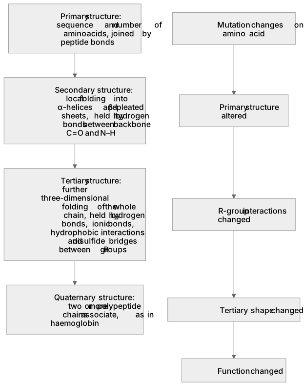
Proteins are built from amino acids joined by peptide bonds, and their shape arises in a hierarchy of levels. The primary structure is the sequence and number of amino acids in the polypeptide chain, held by covalent peptide bonds. The secondary structure is the local folding of the backbone into α-helices and β-pleated sheets, stabilised by hydrogen bonds between the C=O of one amino acid and the N–H of another. The tertiary structure is the further three-dimensional folding of the whole chain, held by hydrogen bonds, ionic bonds, hydrophobic interactions and disulfide bridges between R groups. The quaternary structure exists when two or more polypeptide chains associate, as in haemoglobin. A mutation that changes one amino acid alters the primary structure and can change the tertiary shape and function.
What earns the marks
- Primary structure is the sequence (and number) of amino acids in the polypeptide chain, joined by peptide bonds.
- Secondary structure is the local coiling or folding of the backbone into α-helices and β-pleated sheets, held by hydrogen bonds between backbone C=O and N–H groups.
- Tertiary structure is the further three-dimensional folding of a single polypeptide, stabilised by hydrogen bonds, ionic bonds, hydrophobic interactions and disulfide bridges between R groups.
- Quaternary structure is the association of two or more polypeptide chains (possibly with prosthetic groups) into a functional protein, as in haemoglobin with four chains.
- A change in primary structure, such as a single substituted amino acid, can alter the folding and therefore the tertiary structure and function of the protein.
Common mistakes and how to put them right
Confusing secondary and tertiary structure.
Put it right: Secondary refers to local α-helix and β-pleated sheet folding held by backbone hydrogen bonds, whereas tertiary is the overall 3D fold of the whole chain held by R-group interactions.
Stating that quaternary structure applies to any folded protein.
Put it right: It requires two or more separate polypeptide chains associating, so a single-chain enzyme has no quaternary structure.
Claiming peptide bonds hold the tertiary structure.
Put it right: Peptide bonds form the primary sequence, while tertiary folding depends on interactions between R groups.
Exam tips
- Learn each level with its bonds: peptide bonds for primary, backbone hydrogen bonds for secondary, R-group interactions for tertiary, and chain association for quaternary.
- Use haemoglobin as a concrete quaternary example and a single-chain enzyme such as lysozyme as a protein with no quaternary structure.
- When a question gives a mutation, trace the effect in order: changed amino acid sequence, altered R-group interactions, changed tertiary shape, changed function.
Try the lesson
The start of the Biological molecules: (m) the levels of protein structure lesson, made from checked teaching notes for this specification point.
Have a go first
Haemoglobin is made of four polypeptide chains assembled together. Which level of protein structure does this assembly represent?
Listen
Proteins are built from amino acids joined by peptide bonds, and their shape arises in a hierarchy of levels. The primary structure is the sequence and number of amino acids in the polypeptide chain, held by covalent peptide bonds. The secondary structure is the local folding of the backbone into α-helices and β-pleated sheets, stabilised by hydrogen bonds between the C=O of one amino acid and the N–H of another. The tertiary structure is the further three-dimensional folding of the whole chain, held by hydrogen bonds, ionic bonds, hydrophobic interactions and disulfide bridges between R groups. The quaternary structure exists when two or more polypeptide chains associate, as in haemoglobin. A mutation that changes one amino acid alters the primary structure and can change the tertiary shape and function. A strong answer earns marks for these points. Primary structure is the sequence (and number) of amino acids in the polypeptide chain, joined by peptide bonds. Secondary structure is the local coiling or folding of the backbone into α-helices and β-pleated sheets, held by hydrogen bonds between backbone C=O and N–H groups. Tertiary structure is the further three-dimensional folding of a single polypeptide, stabilised by hydrogen bonds, ionic bonds, hydrophobic interactions and disulfide bridges between R groups. Quaternary structure is the association of two or more polypeptide chains (possibly with prosthetic groups) into a functional protein, as in haemoglobin with four chains. Exam tip: Learn each level with its bonds: peptide bonds for primary, backbone hydrogen bonds for secondary, R-group interactions for tertiary, and chain association for quaternary.
See it
Key words
Say what each word means before you turn the card.
Hierarchy of protein structure
Try answering aloud or in your head before turning the card.
Bonding and R-group interactions
Try answering aloud or in your head before turning the card.
Relationship between sequence and shape
Try answering aloud or in your head before turning the card.
Shape determines function
Try answering aloud or in your head before turning the card.
See every definition
- Hierarchy of protein structure
- The levels in which the shape of a protein arises, from the primary sequence through secondary and tertiary folding to quaternary association of chains.
- Bonding and R-group interactions
- Peptide bonds hold the primary sequence, hydrogen bonds between backbone C=O and N–H groups hold secondary structure, and hydrogen bonds, ionic bonds, hydrophobic interactions and disulfide bridges between R groups hold tertiary structure.
- Relationship between sequence and shape
- A mutation that changes one amino acid alters the primary structure and can change the tertiary shape.
- Shape determines function
- A change in primary structure can alter the folding and therefore the tertiary structure and function of the protein.
Label the diagram
Some boxes have lost their names. Pick the right name for each number.
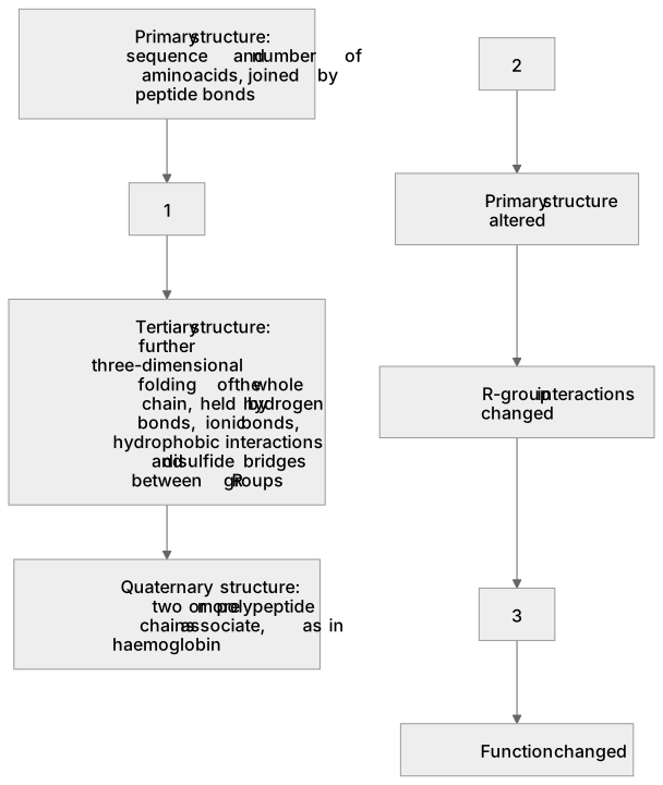
1Box 1
2Box 2
3Box 3
There is more to this lesson
The full lesson goes on to check your understanding and help you earn the marks. Create a free account to carry on, with your progress saved.
A week of Premium from your first lesson · Then the free plan continues · No card, no charge
Test your understanding of Biological molecules: (m) the levels of protein structure
Active recall beats re-reading. Take an instant quiz now to test what you’ve learned and lock it into long-term memory.
A week of Premium from your first lesson · Then the free plan continues · No card, no charge