OCR · A-Level · Biology
Course version: H420 · Version from Sept 2025
(h) photomicrographs of cellular components in a range of eukaryotic cells
Full specification wording
Cell structure: (h) photomicrographs of cellular components in a range of eukaryotic cells
Photomicrographs are images taken through a microscope, and interpreting them is a core practical skill.
Checked against the OCR H420 specification (opens in a new tab) on .
- Point 7 of 10
- 4 key terms
Study notes
From the checked teaching notes for this specification point.
The idea
Photomicrographs are images taken through a microscope, and interpreting them is a core practical skill. You must recognise cellular components across a range of eukaryotic cells, not just one example. Learn to identify the nucleus (often large, with a darker nucleolus), mitochondria (oval with folded internal membranes), chloroplasts (green, stacked thylakoids), the cell surface membrane, a large permanent vacuole in plant cells, the cellulose cell wall, rough and smooth endoplasmic reticulum, Golgi apparatus and ribosomes. Use scale and magnification to judge real size, and compare animal, plant and fungal cells. Distinguish structures by shape, position, membrane pattern and staining, and note that resolution limits what can be seen.
What earns the marks
- Recognises the nucleus and nucleolus in a photomicrograph by its large size, boundary and darker internal region.
- Identifies mitochondria by their oval profile and internal folded membranes (cristae) where resolution allows.
- Identifies chloroplasts in plant cells by their green colour and internal stacked thylakoid membranes.
- Distinguishes a plant cell from an animal cell using the cellulose cell wall and large permanent vacuole.
- Uses the stated magnification or scale bar to estimate the actual size of a named organelle.
- Recognises that some organelles (for example ribosomes) may not be resolvable at the stated magnification.
Common mistakes and how to put them right
Assuming every round structure is a nucleus.
Put it right: Is to check for a boundary and darker nucleolus and to compare with the scale.
Treating a photomicrograph of one cell type as representative of all eukaryotes.
Put it right: Is to compare animal, plant and fungal examples.
Ignoring the scale bar and guessing size.
Put it right: Is to measure the image and divide by magnification to give actual size.
Exam tips
- Read the magnification and scale bar before identifying any structure.
- Match each structure to a named function to confirm your identification.
- Eliminate options that describe structures not visible at the stated resolution.
Try the lesson
The start of the Cell structure: (h) photomicrographs of cellular components in a range of eukaryotic cells lesson, made from checked teaching notes for this specification point.
Have a go first
In a transmission electron micrograph, which feature identifies a mitochondrion?
Listen
Photomicrographs are images taken through a microscope, and interpreting them is a core practical skill. You must recognise cellular components across a range of eukaryotic cells, not just one example. Learn to identify the nucleus (often large, with a darker nucleolus), mitochondria (oval with folded internal membranes), chloroplasts (green, stacked thylakoids), the cell surface membrane, a large permanent vacuole in plant cells, the cellulose cell wall, rough and smooth endoplasmic reticulum, Golgi apparatus and ribosomes. Use scale and magnification to judge real size, and compare animal, plant and fungal cells. Distinguish structures by shape, position, membrane pattern and staining, and note that resolution limits what can be seen. A strong answer earns marks for these points. Recognises the nucleus and nucleolus in a photomicrograph by its large size, boundary and darker internal region. Identifies mitochondria by their oval profile and internal folded membranes (cristae) where resolution allows. Identifies chloroplasts in plant cells by their green colour and internal stacked thylakoid membranes. Distinguishes a plant cell from an animal cell using the cellulose cell wall and large permanent vacuole. A common slip: Assuming every round structure is a nucleus. Instead, is to check for a boundary and darker nucleolus and to compare with the scale. Exam tip: Read the magnification and scale bar before identifying any structure.
See it
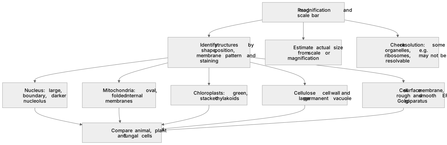
Key words
Say what each word means before you turn the card.
Photomicrograph
Try answering aloud or in your head before turning the card.
Magnification and actual size
Try answering aloud or in your head before turning the card.
Resolution limits of microscopy
Try answering aloud or in your head before turning the card.
Eukaryotic cell diversity
Try answering aloud or in your head before turning the card.
See every definition
- Photomicrograph
- An image taken through a microscope, which you must interpret by recognising cellular components across a range of eukaryotic cells.
- Magnification and actual size
- Using the stated magnification or scale bar to estimate the actual size of a named organelle.
- Resolution limits of microscopy
- Resolution limits what can be seen, so some organelles may not be resolvable at the stated magnification.
- Eukaryotic cell diversity
- Recognising cellular components across a range of eukaryotic cells, not just one example, and comparing animal, plant and fungal cells.
Label the diagram
Some boxes have lost their names. Pick the right name for each number.
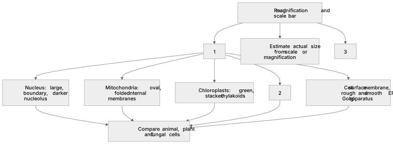
1Box 1
2Box 2
3Box 3
There is more to this lesson
The full lesson goes on to check your understanding and help you earn the marks. Create a free account to carry on, with your progress saved.
A week of Premium from your first lesson · Then the free plan continues · No card, no charge
Test your understanding of Cell structure: (h) photomicrographs of cellular components in a range of eukaryotic cells
Active recall beats re-reading. Take an instant quiz now to test what you’ve learned and lock it into long-term memory.
A week of Premium from your first lesson · Then the free plan continues · No card, no charge