OCR · A-Level · Biology
Course version: H420 · Version from Sept 2025
Cell structure: (j) the importance of the cytoskeleton
The cytoskeleton is a network of protein fibres running through the cytoplasm of eukaryotic cells.
Checked against the OCR H420 specification (opens in a new tab) on .
- Point 9 of 10
- 3 key terms
Study notes
From the checked teaching notes for this specification point.
The idea
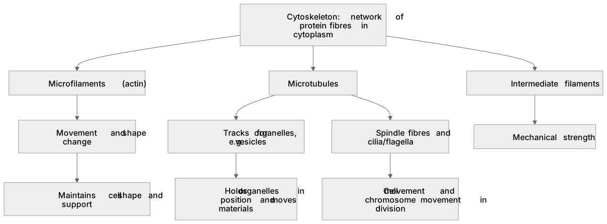
The cytoskeleton is a network of protein fibres running through the cytoplasm of eukaryotic cells. It is not a single structure but several systems working together. Microfilaments (actin) are thin and allow movement and shape change. Microtubules are hollow tubes that form tracks for organelles and make up spindle fibres and cilia/flagella. Intermediate filaments give mechanical strength. Its importance lies in four linked roles: maintaining cell shape and mechanical support; holding organelles in position within the cytoplasm; enabling movement of materials inside the cell, for example vesicles travelling along microtubules; and producing whole-cell movement or movement of cell extensions such as cilia. It also moves chromosomes during cell division by forming the spindle.
What earns the marks
- Identifies the cytoskeleton as a network of protein fibres (microfilaments, microtubules, intermediate filaments) in the cytoplasm.
- States that it maintains cell shape and provides mechanical support to the cell.
- States that it holds organelles in position within the cytoplasm.
- States that it enables movement of materials, for example vesicles moving along microtubules.
- States that it enables cell movement or movement of cilia/flagella.
- States that it forms spindle fibres that move chromosomes during cell division.
Common mistakes and how to put them right
Treating the cytoskeleton as a single structure rather than several protein fibre systems.
Put it right by naming microfilaments, microtubules and intermediate filaments.
Claiming the cytoskeleton is only for movement.
Put it right by including shape maintenance, organelle positioning and support.
Confusing the cytoskeleton with the cell wall.
Put it right by noting the cytoskeleton is internal protein fibres, not a rigid external wall.
Exam tips
- Link each function to a named fibre type and a concrete example, such as vesicles on microtubules.
- Use precise terms: microfilaments, microtubules, spindle fibres, cilia.
- When a question asks for 'importance', give functions rather than just describing structure.
Try the lesson
The start of the Cell structure: (j) the importance of the cytoskeleton lesson, made from checked teaching notes for this specification point.
Have a go first
Which structures make up the cytoskeleton of a eukaryotic cell?
Listen
The cytoskeleton is a network of protein fibres running through the cytoplasm of eukaryotic cells. It is not a single structure but several systems working together. Microfilaments (actin) are thin and allow movement and shape change. Microtubules are hollow tubes that form tracks for organelles and make up spindle fibres and cilia/flagella. Intermediate filaments give mechanical strength. Its importance lies in four linked roles: maintaining cell shape and mechanical support; holding organelles in position within the cytoplasm; enabling movement of materials inside the cell, for example vesicles travelling along microtubules; and producing whole-cell movement or movement of cell extensions such as cilia. It also moves chromosomes during cell division by forming the spindle. A strong answer earns marks for these points. Identifies the cytoskeleton as a network of protein fibres (microfilaments, microtubules, intermediate filaments) in the cytoplasm. States that it maintains cell shape and provides mechanical support to the cell. States that it holds organelles in position within the cytoplasm. States that it enables movement of materials, for example vesicles moving along microtubules. A common slip: Treating the cytoskeleton as a single structure rather than several protein fibre systems. Instead, naming microfilaments, microtubules and intermediate filaments. Exam tip: Link each function to a named fibre type and a concrete example, such as vesicles on microtubules.
See it
Watch it explained
What is the one idea the video explains? Say it in a sentence.
Read the described scene instead
Inside eukaryotic cells, the cytoplasm contains a network of protein fibres called the cytoskeleton. It is not a single structure, but several systems working together. This lesson looks at what each system is made of, and why the cytoskeleton matters to the cell. Three fibre systems make up the cytoskeleton. Microfilaments are made of actin; they are thin and allow movement and changes in shape. Microtubules are hollow tubes that form tracks for organelles, and they make up spindle fibres and cilia and flagella. Intermediate filaments give the cell mechanical strength. Its importance lies in four linked roles. First, maintaining cell shape and giving mechanical support. Second, holding organelles in position within the cytoplasm. These two roles show the cytoskeleton as a supporting framework inside the cell, rather than something used only for movement. Third, the cytoskeleton enables movement of materials inside the cell. Vesicles travel along microtubules, which act as tracks. Fourth, it produces whole-cell movement, or movement of cell extensions such as cilia and flagella. Here is how to turn these functions into a precise answer. Pause and think: which fibre type moves chromosomes, and what is it called during division? Microtubules form the spindle fibres, and the spindle moves chromosomes during cell division. So link each function to a named fibre: microfilaments for shape change, microtubules for vesicles and the spindle, intermediate filaments for strength. Watch for three common errors. Treating the cytoskeleton as a single structure: name microfilaments, microtubules and intermediate filaments instead. Claiming it is only for movement: include shape maintenance, organelle positioning and support. And confusing the cytoskeleton with the cell wall: the cytoskeleton is internal protein fibres, not a rigid external wall. To summarise: the cytoskeleton is a network of protein fibres inside eukaryotic cells, made of microfilaments, microtubules and intermediate filaments. It maintains shape and support, holds organelles in position, moves materials such as vesicles along microtubules, moves the whole cell or its cilia, and forms the spindle that moves chromosomes in division.
Key words
Say what each word means before you turn the card.
Protein fibres
Try answering aloud or in your head before turning the card.
Cell shape and support
Try answering aloud or in your head before turning the card.
Intracellular and whole-cell movement
Try answering aloud or in your head before turning the card.
See every definition
- Protein fibres
- Microfilaments, microtubules and intermediate filaments, which are the systems that make up the cytoskeleton.
- Cell shape and support
- Maintaining cell shape and providing mechanical support, one of the linked roles of the cytoskeleton.
- Intracellular and whole-cell movement
- Movement of materials inside the cell, such as vesicles along microtubules, and movement of the whole cell or cell extensions such as cilia.
Label the diagram
Some boxes have lost their names. Pick the right name for each number.
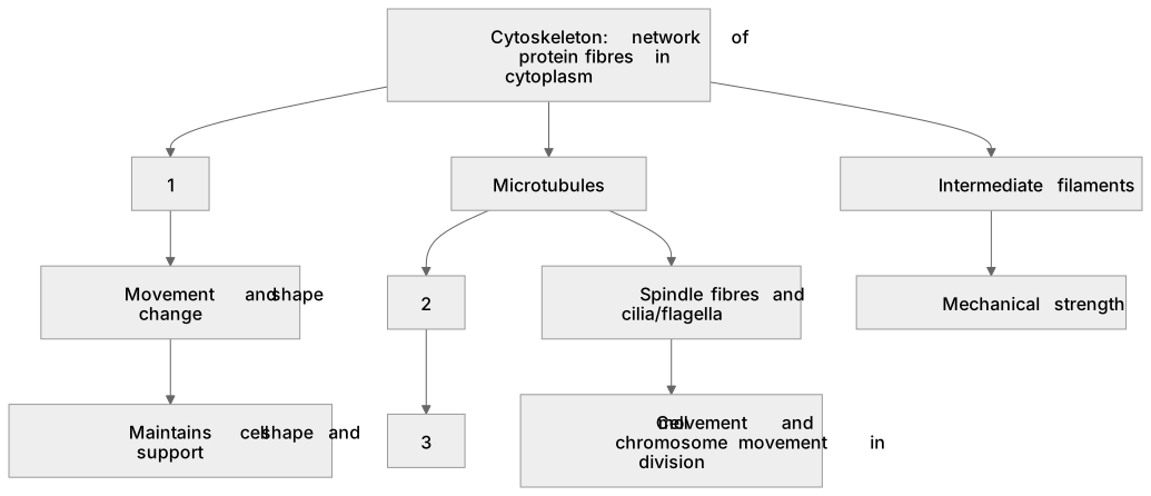
1Box 1
2Box 2
3Box 3
There is more to this lesson
The full lesson goes on to check your understanding and help you earn the marks. Create a free account to carry on, with your progress saved.
A week of Premium from your first lesson · Then the free plan continues · No card, no charge
Test your understanding of Cell structure: (j) the importance of the cytoskeleton
Active recall beats re-reading. Take an instant quiz now to test what you’ve learned and lock it into long-term memory.
A week of Premium from your first lesson · Then the free plan continues · No card, no charge