OCR · A-Level · Biology
Course version: H420 · Version from Sept 2025
(h) the examination of microscope slides to show the histology of exchange…
Full specification wording
Exchange surfaces: (h) the examination of microscope slides to show the histology of exchange surfaces.
This practical involves using a light microscope to examine prepared slides showing the histology of exchange surfaces, such as gill lamellae, alveoli or insect tracheae.
Checked against the OCR H420 specification (opens in a new tab) on .
- Point 7 of 7
- 4 key terms
Study notes
From the checked teaching notes for this specification point.
The idea
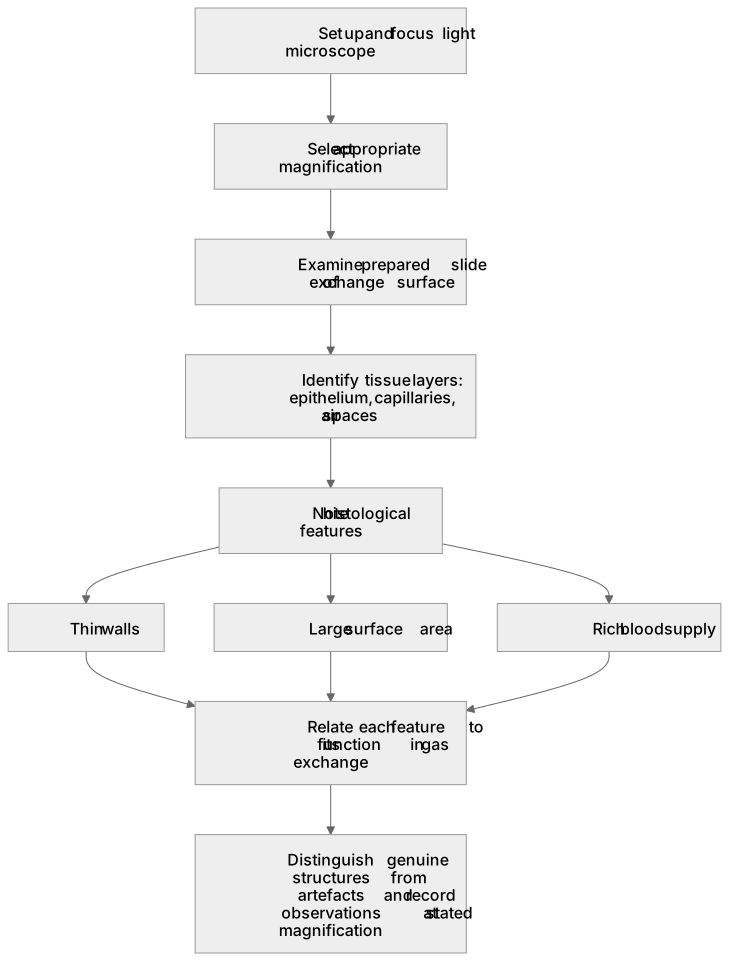
This practical involves using a light microscope to examine prepared slides showing the histology of exchange surfaces, such as gill lamellae, alveoli or insect tracheae. You set up the microscope correctly, select an appropriate magnification, focus the specimen and identify the tissue layers visible. For each slide, note features such as thin walls, extensive blood supply, large surface area and the arrangement of cells that suit efficient gas exchange. You should relate each histological feature to its function, for example a thin epithelium shortening the diffusion distance. Accurate observation means distinguishing real structures from artefacts and recording what is actually seen at the chosen magnification. Assessment checks that you can operate the microscope, identify exchange-surface tissues and explain how their structure supports gas exchange.
What earns the marks
- Sets up and focuses the light microscope correctly, selecting an appropriate magnification to view the tissue.
- Identifies the tissue layers of the exchange surface shown on the slide, such as epithelium, capillaries or air spaces.
- Describes histological features that aid gas exchange, including thin walls, large surface area and a rich blood supply.
- Relates each observed feature to its function in efficient gas exchange.
- Distinguishes genuine structures from artefacts and records observations accurately at the stated magnification.
Common mistakes and how to put them right
Confusing magnification with resolution.
Put it right: Is to recognise that magnification enlarges the image while resolution distinguishes fine detail.
Assuming every dark mark is a structure.
Put it right: Is to distinguish artefacts from genuine tissue features.
Describing features without linking them to function.
Put it right: Is to explain how each feature aids gas exchange.
Exam tips
- Focus at low power first, then increase magnification to examine detail.
- Record the magnification used and describe only what is visible at that magnification.
- Link each histological feature directly to its role in gas exchange.
Try the lesson
The start of the Exchange surfaces: (h) the examination of microscope slides to show the histology of exchange surfaces. lesson, made from checked teaching notes for this specification point.
Have a go first
A student examines a prepared slide of mammalian lung tissue under a light microscope. Which histological feature would indicate that the tissue is from the alveolar region?
Listen
This practical involves using a light microscope to examine prepared slides showing the histology of exchange surfaces, such as gill lamellae, alveoli or insect tracheae. You set up the microscope correctly, select an appropriate magnification, focus the specimen and identify the tissue layers visible. For each slide, note features such as thin walls, extensive blood supply, large surface area and the arrangement of cells that suit efficient gas exchange. You should relate each histological feature to its function, for example a thin epithelium shortening the diffusion distance. Accurate observation means distinguishing real structures from artefacts and recording what is actually seen at the chosen magnification. Assessment checks that you can operate the microscope, identify exchange-surface tissues and explain how their structure supports gas exchange. A strong answer earns marks for these points. Sets up and focuses the light microscope correctly, selecting an appropriate magnification to view the tissue. Identifies the tissue layers of the exchange surface shown on the slide, such as epithelium, capillaries or air spaces. Describes histological features that aid gas exchange, including thin walls, large surface area and a rich blood supply. Relates each observed feature to its function in efficient gas exchange. A common slip: Confusing magnification with resolution. Instead, is to recognise that magnification enlarges the image while resolution distinguishes fine detail. Exam tip: Focus at low power first, then increase magnification to examine detail.
See it
Key words
Say what each word means before you turn the card.
Correct use of the light microscope
Try answering aloud or in your head before turning the card.
Histology of exchange surfaces
Try answering aloud or in your head before turning the card.
Structure–function relationships in gas exchange
Try answering aloud or in your head before turning the card.
Accurate observation and recording
Try answering aloud or in your head before turning the card.
See every definition
- Correct use of the light microscope
- Setting up and focusing the light microscope correctly, selecting an appropriate magnification to view the tissue.
- Histology of exchange surfaces
- The tissue layers of the exchange surface shown on the slide, such as epithelium, capillaries or air spaces.
- Structure–function relationships in gas exchange
- Relating each observed histological feature, such as a thin epithelium shortening the diffusion distance, to its function in efficient gas exchange.
- Accurate observation and recording
- Distinguishing genuine structures from artefacts and recording observations accurately at the stated magnification.
Label the diagram
Some boxes have lost their names. Pick the right name for each number.
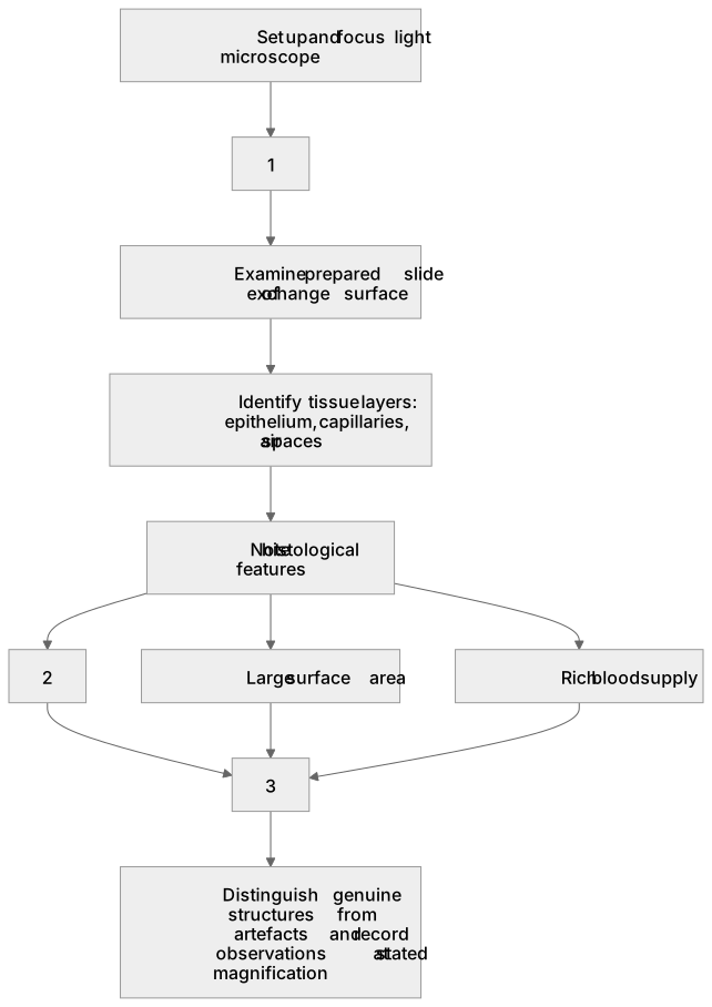
1Box 1
2Box 2
3Box 3
There is more to this lesson
The full lesson goes on to check your understanding and help you earn the marks. Create a free account to carry on, with your progress saved.
A week of Premium from your first lesson · Then the free plan continues · No card, no charge
Test your understanding of Exchange surfaces: (h) the examination of microscope slides to show the histology of exchange surfaces.
Active recall beats re-reading. Take an instant quiz now to test what you’ve learned and lock it into long-term memory.
A week of Premium from your first lesson · Then the free plan continues · No card, no charge