AQA · A-Level · Physical Education
Course version: 7582 · Version from Sept 2016
Cardiovascular system: Myoglobin.
Myoglobin is an oxygen-binding pigment found in skeletal muscle, and it is the muscle's own oxygen store rather than a blood pigment.
Checked against the AQA 7582 specification (opens in a new tab) on .
- Point 14 of 25
- 4 key terms
Study notes
From the checked teaching notes for this specification point.
The idea
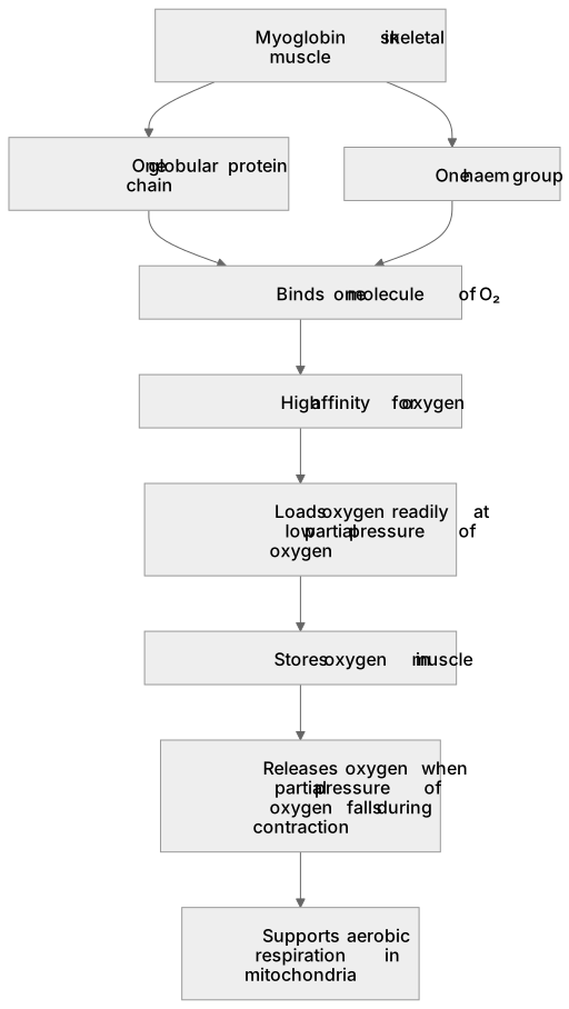
Myoglobin is an oxygen-binding pigment found in skeletal muscle, and it is the muscle's own oxygen store rather than a blood pigment. Structurally it resembles a single subunit of haemoglobin: one globular polypeptide chain wrapped around one haem group, so each myoglobin molecule binds one molecule of oxygen (O₂). It has a very high affinity for oxygen, so it loads oxygen readily even when the partial pressure of oxygen is low, which is exactly the condition inside vigorously contracting muscle. During exercise, muscle oxygen demand rises and blood flow to the working fibres may briefly lag behind, so myoglobin releases its stored oxygen to the mitochondria for aerobic respiration. It also gives slow-twitch fibres their red appearance.
What earns the marks
- Myoglobin is an oxygen-binding pigment located in skeletal muscle (sarcoplasm), not in red blood cells.
- The oxygen it releases supports aerobic respiration in the mitochondria of working muscle fibres.
Common mistakes and how to put them right
Thinking myoglobin is found in red blood cells.
Put it right: It is found in skeletal muscle, whereas haemoglobin is the pigment inside red blood cells.
Believing myoglobin carries four oxygen molecules like haemoglobin.
Put it right: Myoglobin has one haem group and therefore binds one molecule of O₂.
Assuming myoglobin releases oxygen only when partial pressure of oxygen is high.
Put it right: Its high affinity means it loads oxygen at low partial pressure and releases it when tissue partial pressure falls further during contraction.
Confusing myoglobin with mitochondria.
Put it right: Myoglobin stores and transports oxygen, while mitochondria use that oxygen in aerobic respiration.
Exam tips
- Read the stem carefully to check whether the question asks about location, structure or function before choosing an option.
- Compare myoglobin with haemoglobin explicitly: one haem group versus four, muscle versus red blood cell, higher versus lower affinity.
- Link myoglobin to the dissociation curve by noting that its curve sits to the left of the haemoglobin curve at the same partial pressure of oxygen.
- Eliminate options that place myoglobin in blood plasma or describe it as an enzyme rather than an oxygen-binding pigment.
Try the lesson
The start of the Cardiovascular system: Myoglobin. lesson, made from checked teaching notes for this specification point.
Have a go first
In which type of tissue is myoglobin mainly found?
Listen
Myoglobin is an oxygen-binding pigment found in skeletal muscle, and it is the muscle's own oxygen store rather than a blood pigment. Structurally it resembles a single subunit of haemoglobin: one globular polypeptide chain wrapped around one haem group, so each myoglobin molecule binds one molecule of oxygen (O₂). It has a very high affinity for oxygen, so it loads oxygen readily even when the partial pressure of oxygen is low, which is exactly the condition inside vigorously contracting muscle. During exercise, muscle oxygen demand rises and blood flow to the working fibres may briefly lag behind, so myoglobin releases its stored oxygen to the mitochondria for aerobic respiration. It also gives slow-twitch fibres their red appearance. A strong answer earns marks for these points. Myoglobin is an oxygen-binding pigment located in skeletal muscle (sarcoplasm), not in red blood cells. The oxygen it releases supports aerobic respiration in the mitochondria of working muscle fibres. Exam tip: Read the stem carefully to check whether the question asks about location, structure or function before choosing an option.
See it
Key words
Say what each word means before you turn the card.
Myoglobin
Try answering aloud or in your head before turning the card.
Structure of myoglobin
Try answering aloud or in your head before turning the card.
Role of myoglobin during exercise
Try answering aloud or in your head before turning the card.
Affinity for oxygen
Try answering aloud or in your head before turning the card.
See every definition
- Myoglobin
- An oxygen-binding pigment found in skeletal muscle that acts as the muscle's own oxygen store rather than a blood pigment.
- Structure of myoglobin
- One globular polypeptide chain wrapped around one haem group, so each molecule binds one molecule of oxygen.
- Role of myoglobin during exercise
- It releases its stored oxygen to the mitochondria for aerobic respiration when muscle oxygen demand rises and blood flow may briefly lag behind.
- Affinity for oxygen
- A very high affinity means it loads oxygen readily even when the partial pressure of oxygen is low, as inside vigorously contracting muscle.
Label the diagram
Some boxes have lost their names. Pick the right name for each number.
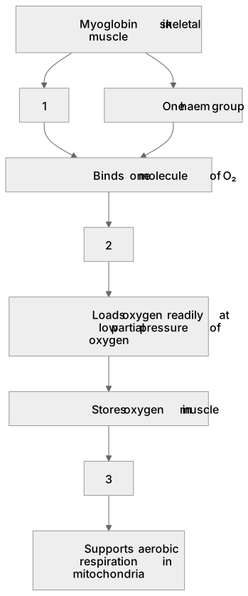
1Box 1
2Box 2
3Box 3
There is more to this lesson
The full lesson goes on to check your understanding and help you earn the marks. Create a free account to carry on, with your progress saved.
A week of Premium from your first lesson · Then the free plan continues · No card, no charge
Test your understanding of Cardiovascular system: Myoglobin.
Active recall beats re-reading. Take an instant quiz now to test what you’ve learned and lock it into long-term memory.
A week of Premium from your first lesson · Then the free plan continues · No card, no charge