AQA · A-Level · Physical Education
Course version: 7582 · Version from Sept 2016
Neuromuscular system: Muscle spindles.
Muscle spindles are proprioceptors located within skeletal muscle.
Checked against the AQA 7582 specification (opens in a new tab) on .
- Point 9 of 16
- 4 key terms
Study notes
From the checked teaching notes for this specification point.
The idea
Muscle spindles are proprioceptors located within skeletal muscle. They detect changes in muscle length and the rate of that change. When a muscle is stretched rapidly, the spindle sends sensory signals to the central nervous system, triggering a stretch reflex: the stretched muscle contracts to resist the stretch. This reflex maintains posture and protects against overstretching. In sport, plyometric training utilises this stretch reflex to produce a more powerful concentric contraction following a rapid eccentric stretch. In contrast, Proprioceptive Neuromuscular Facilitation (PNF) stretching relies on Golgi tendon organs to override the stretch reflex via autogenic inhibition, allowing the muscle to relax and stretch further.
What earns the marks
- Muscle spindles are proprioceptors found in skeletal muscle that detect stretch and rate of change in length.
- The reflex involves sensory neurones sending signals to the spinal cord and motor neurones causing contraction.
- Spindles provide feedback for posture and movement coordination.
- Plyometrics uses the stretch reflex initiated by muscle spindles to generate powerful contractions, whereas PNF stretching overrides it using Golgi tendon organs.
Common mistakes and how to put them right
Confusing muscle spindles with Golgi tendon organs.
Put it right: Spindles detect stretch and cause contraction (stretch reflex), while Golgi tendon organs detect tension and cause relaxation (autogenic inhibition).
Thinking PNF stretching relies on muscle spindles.
Put it right: PNF actually uses Golgi tendon organs to override the spindle's stretch reflex.
Believing the stretch reflex is a voluntary response.
Put it right: It is an involuntary reflex initiated by the spinal cord.
Exam tips
- Remember the sequence: stretch detected by spindle → sensory neurone → spinal cord → motor neurone → muscle contraction.
- Link muscle spindles to the stretch reflex and plyometric training, not to the relaxation phase of PNF.
- Use the term 'stretch reflex' accurately and link it to protection against overstretching.
Try the lesson
The start of the Neuromuscular system: Muscle spindles. lesson, made from checked teaching notes for this specification point.
Have a go first
Which type of receptor detects changes in the length of a skeletal muscle?
Listen
Muscle spindles are proprioceptors located within skeletal muscle. They detect changes in muscle length and the rate of that change. When a muscle is stretched rapidly, the spindle sends sensory signals to the central nervous system, triggering a stretch reflex: the stretched muscle contracts to resist the stretch. This reflex maintains posture and protects against overstretching. In sport, plyometric training utilises this stretch reflex to produce a more powerful concentric contraction following a rapid eccentric stretch. In contrast, Proprioceptive Neuromuscular Facilitation (PNF) stretching relies on Golgi tendon organs to override the stretch reflex via autogenic inhibition, allowing the muscle to relax and stretch further. A strong answer earns marks for these points. Muscle spindles are proprioceptors found in skeletal muscle that detect stretch and rate of change in length. The reflex involves sensory neurones sending signals to the spinal cord and motor neurones causing contraction. Spindles provide feedback for posture and movement coordination. Plyometrics uses the stretch reflex initiated by muscle spindles to generate powerful contractions, whereas PNF stretching overrides it using Golgi tendon organs. Exam tip: Remember the sequence: stretch detected by spindle → sensory neurone → spinal cord → motor neurone → muscle contraction.
See it
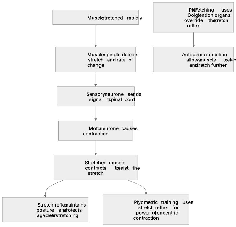
Key words
Say what each word means before you turn the card.
Muscle spindles
Try answering aloud or in your head before turning the card.
Stretch reflex
Try answering aloud or in your head before turning the card.
Plyometric training
Try answering aloud or in your head before turning the card.
Proprioceptive Neuromuscular Facilitation (PNF) stretching
Try answering aloud or in your head before turning the card.
See every definition
- Muscle spindles
- Proprioceptors located within skeletal muscle that detect changes in muscle length and the rate of that change.
- Stretch reflex
- When a muscle is stretched rapidly, the spindle sends sensory signals to the central nervous system, triggering the stretched muscle to contract to resist the stretch.
- Plyometric training
- Training that utilises the stretch reflex to produce a more powerful concentric contraction following a rapid eccentric stretch.
- Proprioceptive Neuromuscular Facilitation (PNF) stretching
- Stretching that relies on Golgi tendon organs to override the stretch reflex via autogenic inhibition, allowing the muscle to relax and stretch further.
Label the diagram
Some boxes have lost their names. Pick the right name for each number.
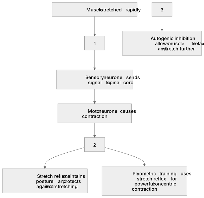
1Box 1
2Box 2
3Box 3
There is more to this lesson
The full lesson goes on to check your understanding and help you earn the marks. Create a free account to carry on, with your progress saved.
A week of Premium from your first lesson · Then the free plan continues · No card, no charge
Test your understanding of Neuromuscular system: Muscle spindles.
Active recall beats re-reading. Take an instant quiz now to test what you’ve learned and lock it into long-term memory.
A week of Premium from your first lesson · Then the free plan continues · No card, no charge