OCR · A-Level · Physics
Course version: H556 · Version from Sept 2015
(d) positron emission tomography (PET) scanner; annihilation of…
Full specification wording
Diagnostic methods in medicine: (d) positron emission tomography (PET) scanner; annihilation of positron–electron pairs; formation of image
A PET scanner images the distribution of a positron-emitting radionuclide, often fluorine-18 in fluorodeoxyglucose, which is taken up by metabolically active cells.
Checked against the OCR H556 specification (opens in a new tab) on .
- Point 4 of 5
- 4 key terms
Study notes
From the checked teaching notes for this specification point.
The idea
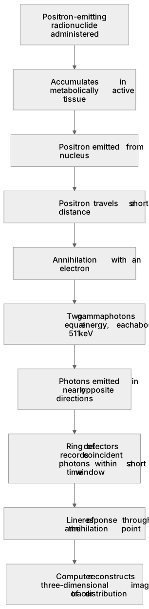
A PET scanner images the distribution of a positron-emitting radionuclide, often fluorine-18 in fluorodeoxyglucose, which is taken up by metabolically active cells. When a positron is emitted, it travels a short distance and annihilates with an electron. The annihilation converts the total rest mass into two gamma photons of equal energy, each about 511 keV, emitted in almost opposite directions. A ring of detectors surrounds the patient. When two detectors on opposite sides register photons within a very short coincidence time window, the annihilation event is assumed to lie on the line joining them. The computer collects many such coincidence events and reconstructs a three-dimensional image of tracer concentration. The technique gives functional information about metabolism and is often combined with CT for anatomical reference.
What earns the marks
- A positron-emitting radionuclide is administered and accumulates in metabolically active tissue.
- A positron emitted from the nucleus travels a short distance before annihilating with an electron.
- Annihilation converts mass into two gamma photons of equal energy, each about 511 keV, emitted in nearly opposite directions.
- A ring of detectors records coincident gamma photons within a short time window, defining a line of response through the annihilation point.
- The computer reconstructs a three-dimensional image of tracer distribution from many coincidence events.
Common mistakes and how to put them right
Thinking the positron itself is detected.
Put it right: The positron annihilates and the two gamma photons are detected.
Believing the two gamma photons have different energies.
Put it right: They each have about 511 keV, equal to the electron rest energy.
Assuming the photons are emitted exactly 180° apart in every case.
Put it right: They are emitted in nearly opposite directions, and small deviations affect resolution.
Confusing PET with gamma camera imaging.
Put it right: PET detects coincidence gamma pairs from annihilation, while a gamma camera detects single gamma photons with a collimator.
Exam tips
- State the annihilation process clearly: positron plus electron gives two gamma photons of about 511 keV each.
- Explain coincidence detection: two detectors on opposite sides must register photons within a very short time window.
- Link the line of response to the reconstruction of a three-dimensional image.
Try the lesson
The start of the Diagnostic methods in medicine: (d) positron emission tomography (PET) scanner; annihilation of positron–electron pairs; formation of image lesson, made from checked teaching notes for this specification point.
Listen
A PET scanner images the distribution of a positron-emitting radionuclide, often fluorine-18 in fluorodeoxyglucose, which is taken up by metabolically active cells. When a positron is emitted, it travels a short distance and annihilates with an electron. The annihilation converts the total rest mass into two gamma photons of equal energy, each about 511 keV, emitted in almost opposite directions. A ring of detectors surrounds the patient. When two detectors on opposite sides register photons within a very short coincidence time window, the annihilation event is assumed to lie on the line joining them. The computer collects many such coincidence events and reconstructs a three-dimensional image of tracer concentration. The technique gives functional information about metabolism and is often combined with CT for anatomical reference. A strong answer earns marks for these points. A positron-emitting radionuclide is administered and accumulates in metabolically active tissue. A positron emitted from the nucleus travels a short distance before annihilating with an electron. Annihilation converts mass into two gamma photons of equal energy, each about 511 keV, emitted in nearly opposite directions. A ring of detectors records coincident gamma photons within a short time window, defining a line of response through the annihilation point. Exam tip: State the annihilation process clearly: positron plus electron gives two gamma photons of about 511 keV each.
See it
Key words
Say what each word means before you turn the card.
Annihilation
Try answering aloud or in your head before turning the card.
Coincidence detection
Try answering aloud or in your head before turning the card.
Three-dimensional image reconstruction
Try answering aloud or in your head before turning the card.
Positron-emitting radionuclide
Try answering aloud or in your head before turning the card.
See every definition
- Annihilation
- The process in which a positron and an electron convert the total rest mass into two gamma photons of equal energy, each about 511 keV, emitted in almost opposite directions.
- Coincidence detection
- Two detectors on opposite sides of a ring surrounding the patient register photons within a very short coincidence time window, so the annihilation event is assumed to lie on the line joining them.
- Three-dimensional image reconstruction
- The computer collects many coincidence events and reconstructs a three-dimensional image of tracer concentration.
- Positron-emitting radionuclide
- A substance, often fluorine-18 in fluorodeoxyglucose, that is administered and taken up by metabolically active cells, emitting a positron that travels a short distance before annihilating with an electron.
Label the diagram
Some boxes have lost their names. Pick the right name for each number.
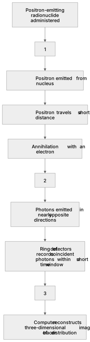
1Box 1
2Box 2
3Box 3
There is more to this lesson
The full lesson goes on to check your understanding and help you earn the marks. Create a free account to carry on, with your progress saved.
A week of Premium from your first lesson · Then the free plan continues · No card, no charge
Test your understanding of Diagnostic methods in medicine: (d) positron emission tomography (PET) scanner; annihilation of positron–electron pairs; formation of image
Active recall beats re-reading. Take an instant quiz now to test what you’ve learned and lock it into long-term memory.
A week of Premium from your first lesson · Then the free plan continues · No card, no charge