OCR · A-Level · Physics
Course version: H556 · Version from Sept 2015
Diagnostic methods in medicine: (c) diagnosis using gamma camera
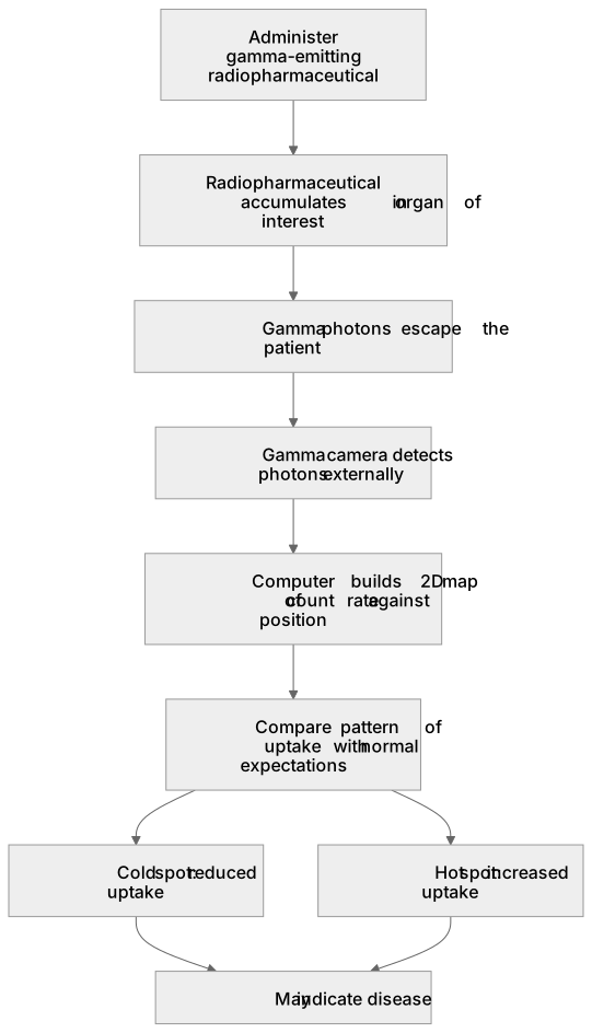
Diagnosis with a gamma camera begins by administering a radiopharmaceutical whose gamma-emitting radionuclide is chosen to accumulate in the organ of interest.
Checked against the OCR H556 specification (opens in a new tab) on .
- Point 3 of 5
- 4 key terms
Study notes
From the checked teaching notes for this specification point.
The idea
Diagnosis with a gamma camera begins by administering a radiopharmaceutical whose gamma-emitting radionuclide is chosen to accumulate in the organ of interest. For example, technetium-99m labelled to a carrier may concentrate in bone or kidneys. Gamma photons escaping the patient are detected by the camera, and the computer builds a two-dimensional map of count rate against position. A healthy organ shows a characteristic pattern of uptake; disease may appear as a cold spot where uptake is reduced, or a hot spot where uptake is increased. The clinician compares the image with normal expectations and may quantify counts in regions of interest. The technique gives functional information about physiology rather than sharp anatomical detail, and the collimator limits sensitivity, so image quality depends on tracer activity, acquisition time and patient positioning.
What earns the marks
- A gamma-emitting radiopharmaceutical is administered and is designed to accumulate in the target organ or tissue.
- Gamma photons emitted from within the patient are detected externally by the gamma camera.
- The computer forms a two-dimensional image showing the distribution of tracer uptake.
- Areas of abnormally high or low uptake are interpreted as hot spots or cold spots that may indicate disease.
- The image provides functional information about the organ, and diagnosis depends on comparing the pattern with normal uptake.
Common mistakes and how to put them right
Thinking the gamma camera itself emits radiation into the patient.
Put it right: The radiation comes from the administered radiopharmaceutical.
Confusing a cold spot with a hot spot.
Put it right: A cold spot has reduced tracer uptake, while a hot spot has increased uptake.
Assuming the gamma camera gives detailed anatomical images like a CT scan.
Put it right: It primarily shows functional distribution of tracer.
Believing any gamma emitter is suitable.
Put it right: The radionuclide must emit gamma rays of suitable energy and have a half-life long enough for imaging but short enough to limit dose.
Exam tips
- State the clinical purpose: to assess organ function by mapping tracer uptake.
- Use the terms hot spot and cold spot correctly and link each to increased or decreased uptake.
- Mention that the choice of radiopharmaceutical depends on the organ being investigated.
Try the lesson
The start of the Diagnostic methods in medicine: (c) diagnosis using gamma camera lesson, made from checked teaching notes for this specification point.
Listen
Diagnosis with a gamma camera begins by administering a radiopharmaceutical whose gamma-emitting radionuclide is chosen to accumulate in the organ of interest. For example, technetium-99m labelled to a carrier may concentrate in bone or kidneys. Gamma photons escaping the patient are detected by the camera, and the computer builds a two-dimensional map of count rate against position. A healthy organ shows a characteristic pattern of uptake; disease may appear as a cold spot where uptake is reduced, or a hot spot where uptake is increased. The clinician compares the image with normal expectations and may quantify counts in regions of interest. The technique gives functional information about physiology rather than sharp anatomical detail, and the collimator limits sensitivity, so image quality depends on tracer activity, acquisition time and patient positioning. A strong answer earns marks for these points. A gamma-emitting radiopharmaceutical is administered and is designed to accumulate in the target organ or tissue. Gamma photons emitted from within the patient are detected externally by the gamma camera. The computer forms a two-dimensional image showing the distribution of tracer uptake. Areas of abnormally high or low uptake are interpreted as hot spots or cold spots that may indicate disease. Exam tip: State the clinical purpose: to assess organ function by mapping tracer uptake.
See it
Key words
Say what each word means before you turn the card.
Functional medical imaging
Try answering aloud or in your head before turning the card.
Radiopharmaceutical uptake and distribution
Try answering aloud or in your head before turning the card.
Interpretation of hot spots and cold spots
Try answering aloud or in your head before turning the card.
Clinical diagnosis from count maps
Try answering aloud or in your head before turning the card.
See every definition
- Functional medical imaging
- A technique that gives functional information about physiology rather than sharp anatomical detail, showing the distribution of tracer uptake.
- Radiopharmaceutical uptake and distribution
- A gamma-emitting radiopharmaceutical is administered and designed to accumulate in the target organ or tissue, and the computer forms a two-dimensional image showing its distribution.
- Interpretation of hot spots and cold spots
- Areas of abnormally high or low uptake are interpreted as hot spots or cold spots that may indicate disease.
- Clinical diagnosis from count maps
- Diagnosis depends on comparing the pattern of uptake with normal expectations, and the clinician may quantify counts in regions of interest.
Label the diagram
Some boxes have lost their names. Pick the right name for each number.
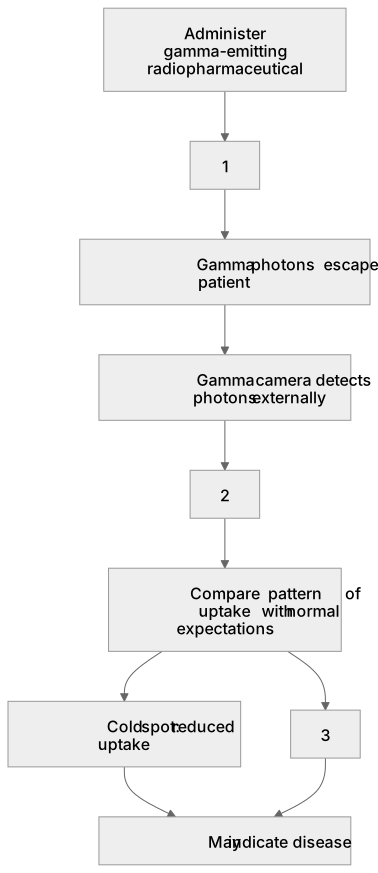
1Box 1
2Box 2
3Box 3
There is more to this lesson
The full lesson goes on to check your understanding and help you earn the marks. Create a free account to carry on, with your progress saved.
A week of Premium from your first lesson · Then the free plan continues · No card, no charge
Test your understanding of Diagnostic methods in medicine: (c) diagnosis using gamma camera
Active recall beats re-reading. Take an instant quiz now to test what you’ve learned and lock it into long-term memory.
A week of Premium from your first lesson · Then the free plan continues · No card, no charge