OCR · A-Level · Physics
Course version: H556 · Version from Sept 2015
(f) computerised axial tomography (CAT) scanning; components – rotating…
Full specification wording
Nuclear fission and fusion: (f) computerised axial tomography (CAT) scanning; components – rotating X-tube producing a thin fan-shaped X-ray beam, ring of detectors, computer software and display
A CAT scanner builds a cross-sectional image by measuring X-ray attenuation from many directions.
Checked against the OCR H556 specification (opens in a new tab) on .
- Point 18 of 18
- 4 key terms
Study notes
From the checked teaching notes for this specification point.
The idea
A CAT scanner builds a cross-sectional image by measuring X-ray attenuation from many directions. The X-tube rotates around the patient and emits a thin fan-shaped X-ray beam that passes through a narrow slice of the body. A ring of detectors opposite the tube measures the transmitted intensity at many angles. Because the beam is thin and the tube rotates, each point in the slice is sampled from multiple directions, so the computer software can reconstruct the attenuation at each point and display a detailed cross-sectional image. The fan shape allows a complete slice to be covered in one rotation, and the ring of detectors collects the transmitted beam efficiently. The result is a two-dimensional map of attenuation, which can reveal small differences between soft tissues.
What earns the marks
- The X-tube rotates around the patient and produces a thin fan-shaped X-ray beam.
- A ring of detectors measures the transmitted X-ray intensity at many angles around the patient.
- The beam is thin so that it examines a narrow slice of the body, giving good spatial resolution.
- Computer software reconstructs the attenuation data into a cross-sectional image.
- The image is displayed on a monitor for interpretation by a radiologist.
Common mistakes and how to put them right
Thinking the detectors rotate while the tube is fixed.
Put it right: In a CAT scanner the X-tube rotates and the ring of detectors surrounds the patient.
Believing the beam is a broad cone that covers the whole body.
Put it right: The beam is a thin fan that examines a narrow slice.
Assuming the computer produces the image directly from a single projection.
Put it right: Many projections from different angles are needed for reconstruction.
Confusing CAT with a simple X-ray photograph.
Put it right: A CAT scan produces a cross-sectional image from measured attenuation data, not a single shadow.
Exam tips
- Name each component and state its function: rotating X-tube, fan-shaped beam, ring of detectors, computer software, display.
- Use the phrase thin fan-shaped beam to emphasise the narrow slice and the coverage of the detector ring.
- Explain that many projections from different angles are combined by software to reconstruct the cross-section.
Try the lesson
The start of the Nuclear fission and fusion: (f) computerised axial tomography (CAT) scanning; components – rotating X-tube producing a thin fan-shaped X-ray beam, ring of detectors, computer software and display lesson, made from checked teaching notes for this specification point.
Listen
A CAT scanner builds a cross-sectional image by measuring X-ray attenuation from many directions. The X-tube rotates around the patient and emits a thin fan-shaped X-ray beam that passes through a narrow slice of the body. A ring of detectors opposite the tube measures the transmitted intensity at many angles. Because the beam is thin and the tube rotates, each point in the slice is sampled from multiple directions, so the computer software can reconstruct the attenuation at each point and display a detailed cross-sectional image. The fan shape allows a complete slice to be covered in one rotation, and the ring of detectors collects the transmitted beam efficiently. The result is a two-dimensional map of attenuation, which can reveal small differences between soft tissues. A strong answer earns marks for these points. The X-tube rotates around the patient and produces a thin fan-shaped X-ray beam. A ring of detectors measures the transmitted X-ray intensity at many angles around the patient. The beam is thin so that it examines a narrow slice of the body, giving good spatial resolution. Computer software reconstructs the attenuation data into a cross-sectional image. Exam tip: Name each component and state its function: rotating X-tube, fan-shaped beam, ring of detectors, computer software, display.
See it
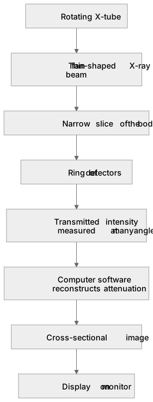
Put it in order
Put these steps in the order they happen.
- 1. Transmitted intensity measured at many angles
- 2. Display on monitor
- 3. Computer software reconstructs attenuation
- 4. Rotating X-tube
- 5. Thin fan-shaped X-ray beam
- 6. Cross-sectional image
- 7. Narrow slice of the body
- 8. Ring of detectors
Key words
Say what each word means before you turn the card.
Rotating X-tube and fan-shaped beam
Try answering aloud or in your head before turning the card.
Ring of detectors and transmitted intensity
Try answering aloud or in your head before turning the card.
Computer reconstruction of attenuation data
Try answering aloud or in your head before turning the card.
Cross-sectional imaging and display
Try answering aloud or in your head before turning the card.
See every definition
- Rotating X-tube and fan-shaped beam
- The X-tube rotates around the patient and produces a thin fan-shaped X-ray beam that passes through a narrow slice of the body.
- Ring of detectors and transmitted intensity
- A ring of detectors surrounds the patient and measures the transmitted X-ray intensity at many angles.
- Computer reconstruction of attenuation data
- Computer software reconstructs the attenuation data into a cross-sectional image, combining many projections from different angles.
- Cross-sectional imaging and display
- The result is a two-dimensional map of attenuation, displayed on a monitor for interpretation by a radiologist.
Label the diagram
Some boxes have lost their names. Pick the right name for each number.
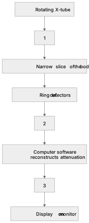
1Box 1
2Box 2
3Box 3
There is more to this lesson
The full lesson goes on to check your understanding and help you earn the marks. Create a free account to carry on, with your progress saved.
A week of Premium from your first lesson · Then the free plan continues · No card, no charge
Test your understanding of Nuclear fission and fusion: (f) computerised axial tomography (CAT) scanning; components – rotating X-tube producing a thin fan-shaped X-ray beam, ring of detectors, computer software and display
Active recall beats re-reading. Take an instant quiz now to test what you’ve learned and lock it into long-term memory.
A week of Premium from your first lesson · Then the free plan continues · No card, no charge