AQA · GCSE · Biology
Course version: 8461 · Version from Sept 2018
This has enabled biologists to see and understand many more sub-cellular…
Full specification wording
Microscopy: This has enabled biologists to see and understand many more sub-cellular structures.
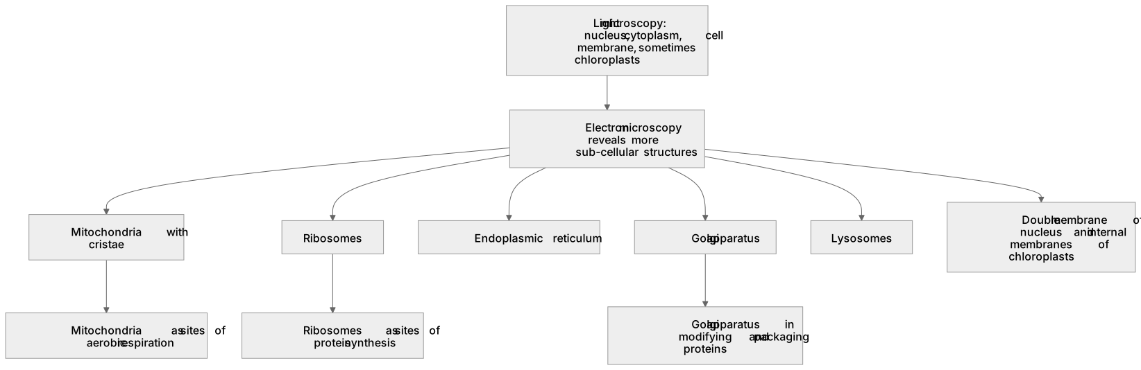
Before electron microscopy, cell biology relied on light microscopes, which resolved the nucleus, cytoplasm, cell membrane and sometimes chloroplasts, but little else.
Checked against the AQA 8461 specification (opens in a new tab) on .
- Point 4 of 4
- 3 key terms
Study notes
From the checked teaching notes for this specification point.
The idea
Before electron microscopy, cell biology relied on light microscopes, which resolved the nucleus, cytoplasm, cell membrane and sometimes chloroplasts, but little else. Electron microscopy revealed organelles such as mitochondria with their cristae, ribosomes, the endoplasmic reticulum, Golgi apparatus and lysosomes, and showed the double membrane of the nucleus and the internal membranes of chloroplasts. Seeing these structures allowed biologists to propose functions: ribosomes as sites of protein synthesis, mitochondria as sites of aerobic respiration and the Golgi apparatus in modifying and packaging proteins. For example, the observation that pancreatic cells are packed with rough endoplasmic reticulum supported the idea that they secrete proteins. Structure therefore informs function, and improved microscopy has driven advances in cell biology, medicine and biotechnology.
What earns the marks
- Electron microscopy revealed organelles that light microscopy could not resolve, including ribosomes, endoplasmic reticulum, Golgi apparatus and lysosomes.
- Observing a structure allows biologists to infer its function, for example ribosomes as sites of protein synthesis.
- Correlating organelle abundance with cell role, such as many mitochondria in muscle cells, supports structure-function reasoning.
- Advances in microscopy have contributed to fields such as medicine, genetics and biotechnology.
- Understanding sub-cellular structure underpins later topics including transport, enzymes and cell division.
Common mistakes and how to put them right
Listing organelles without linking each to the function it performs.
Put it right: Pair every structure with its role, such as ribosomes with protein synthesis.
Implying that light microscopes show no organelles at all.
Put it right: They do show the nucleus, cytoplasm, cell membrane and often chloroplasts, but not finer structures.
Treating the history of microscopy as a list of dates rather than explaining how better resolution led to new biological understanding.
Exam tips
- Use the structure-function link explicitly: name the organelle, then state what its structure allows it to do.
- Choose examples that match the cell type, such as many mitochondria in a sperm cell or muscle cell.
- If asked about evidence, describe what was observed and the conclusion drawn, keeping observation and inference distinct.
Try the lesson
The start of the Microscopy: This has enabled biologists to see and understand many more sub-cellular structures. lesson, made from checked teaching notes for this specification point.
Have a go first
What has the electron microscope enabled biologists to see and understand?
Listen
Before electron microscopy, cell biology relied on light microscopes, which resolved the nucleus, cytoplasm, cell membrane and sometimes chloroplasts, but little else. Electron microscopy revealed organelles such as mitochondria with their cristae, ribosomes, the endoplasmic reticulum, Golgi apparatus and lysosomes, and showed the double membrane of the nucleus and the internal membranes of chloroplasts. Seeing these structures allowed biologists to propose functions: ribosomes as sites of protein synthesis, mitochondria as sites of aerobic respiration and the Golgi apparatus in modifying and packaging proteins. For example, the observation that pancreatic cells are packed with rough endoplasmic reticulum supported the idea that they secrete proteins. Structure therefore informs function, and improved microscopy has driven advances in cell biology, medicine and biotechnology. A strong answer earns marks for these points. Electron microscopy revealed organelles that light microscopy could not resolve, including ribosomes, endoplasmic reticulum, Golgi apparatus and lysosomes. Observing a structure allows biologists to infer its function, for example ribosomes as sites of protein synthesis. Correlating organelle abundance with cell role, such as many mitochondria in muscle cells, supports structure-function reasoning. Advances in microscopy have contributed to fields such as medicine, genetics and biotechnology. Exam tip: Use the structure-function link explicitly: name the organelle, then state what its structure allows it to do.
See it
Key words
Say what each word means before you turn the card.
Discovery of sub-cellular structures
Try answering aloud or in your head before turning the card.
Structure and function of organelles
Try answering aloud or in your head before turning the card.
Impact of technology on biological knowledge
Try answering aloud or in your head before turning the card.
See every definition
- Discovery of sub-cellular structures
- Electron microscopy revealed organelles that light microscopy could not resolve, including ribosomes, endoplasmic reticulum, Golgi apparatus and lysosomes.
- Structure and function of organelles
- Observing a structure allows biologists to infer its function, for example ribosomes as sites of protein synthesis.
- Impact of technology on biological knowledge
- Improved microscopy has driven advances in cell biology, medicine and biotechnology.
Label the diagram
Some boxes have lost their names. Pick the right name for each number.
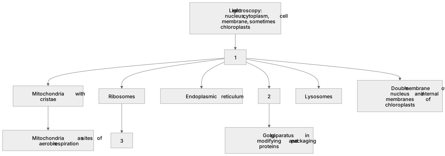
1Box 1
2Box 2
3Box 3
There is more to this lesson
The full lesson goes on to check your understanding and help you earn the marks. Create a free account to carry on, with your progress saved.
A week of Premium from your first lesson · Then the free plan continues · No card, no charge
Test your understanding of Microscopy: This has enabled biologists to see and understand many more sub-cellular structures.
Active recall beats re-reading. Take an instant quiz now to test what you’ve learned and lock it into long-term memory.
A week of Premium from your first lesson · Then the free plan continues · No card, no charge