AQA · GCSE · Biology
Course version: 8461 · Version from Sept 2018
This means that it can be used to study cells in much finer detail.
Full specification wording
Microscopy: This means that it can be used to study cells in much finer detail.
Because electrons have a much shorter wavelength than visible light, an electron microscope resolves points that are only about 0.2 nm apart, whereas a light microscope cannot separate points…
Checked against the AQA 8461 specification (opens in a new tab) on .
- Point 3 of 4
- 4 key terms
Study notes
From the checked teaching notes for this specification point.
The idea
Because electrons have a much shorter wavelength than visible light, an electron microscope resolves points that are only about 0.2 nm apart, whereas a light microscope cannot separate points closer than about 200 nm. This thousand-fold improvement in resolving power means structures that blur together under a light microscope become sharp and separate in an electron micrograph. For instance, a light microscope shows a mitochondrion as a faint oval, but an electron micrograph reveals its double membrane and folded cristae. Similarly, the internal membranes of a chloroplast or the detail of a synapse become visible. Finer detail is therefore a direct consequence of better resolution, not merely of greater magnification, and it allows biologists to relate structure to function more precisely.
What earns the marks
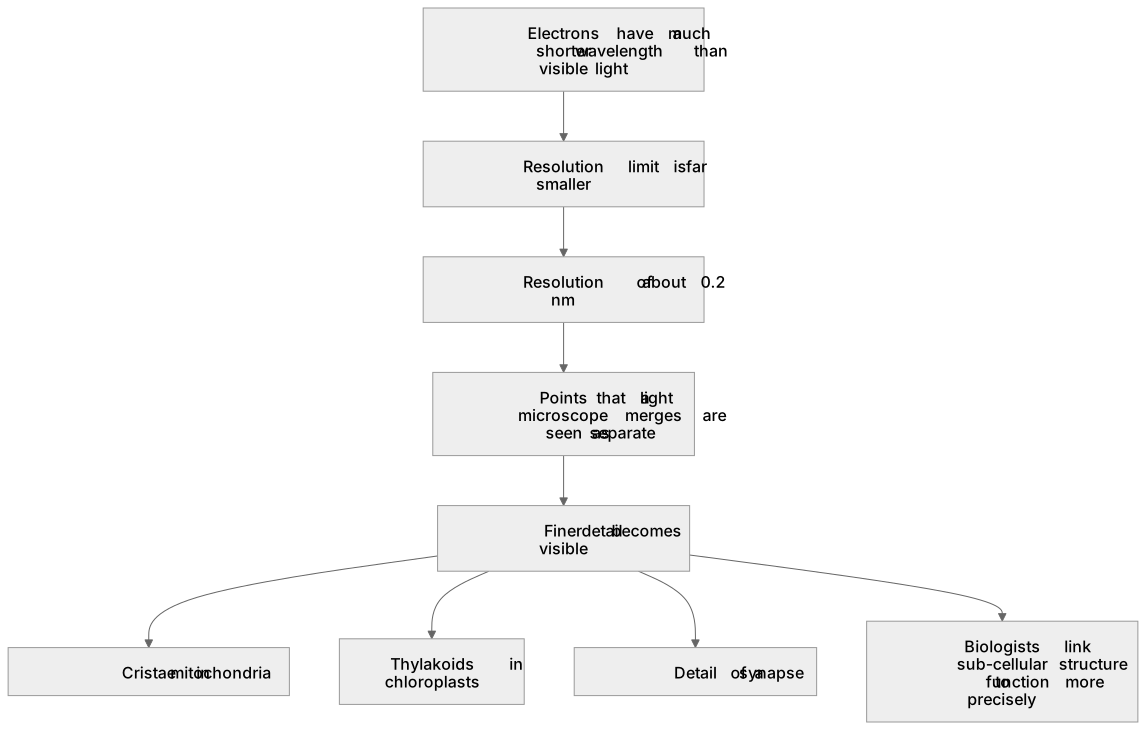
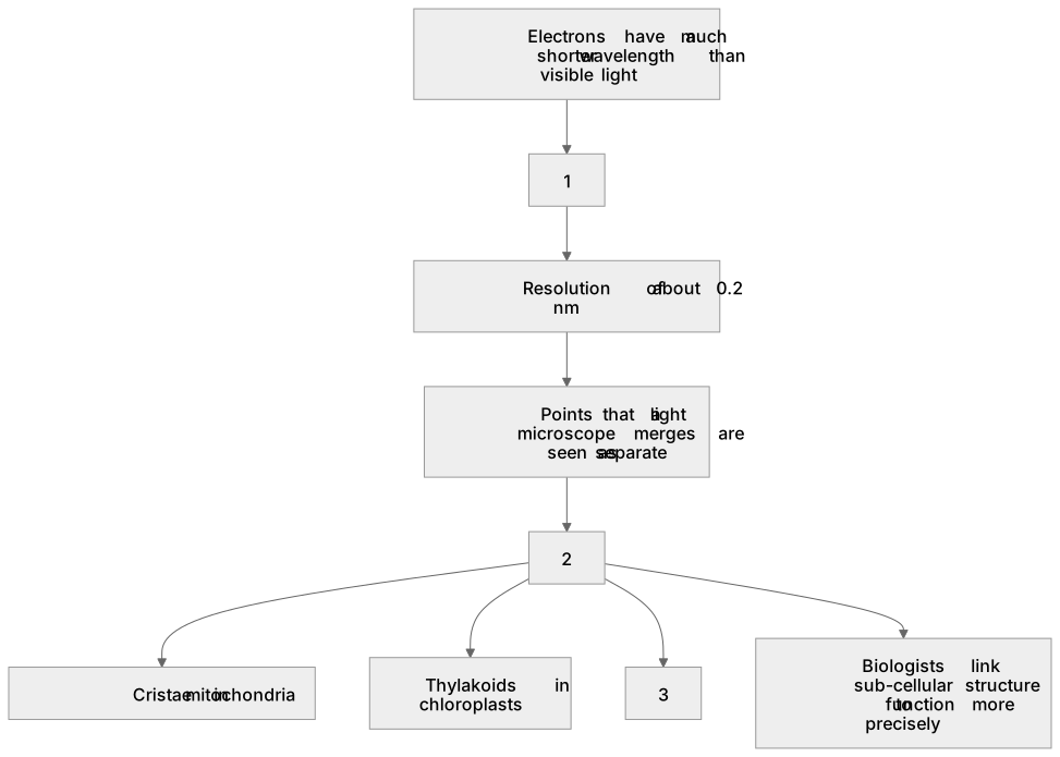
- Electrons have a much shorter wavelength than visible light, so the resolution limit is far smaller.
- Resolution of about 0.2 nm allows points that a light microscope merges to be seen as separate.
- Finer detail includes internal membranes and organelles such as cristae in mitochondria and thylakoids in chloroplasts.
- The improvement follows from resolving power, not from magnification alone, because enlarging a blurred image adds no new information.
- Finer structural detail helps biologists link sub-cellular structure to its function.
- Specimens for electron microscopy are usually dead and dehydrated, which is a limitation when studying living cells.
Common mistakes and how to put them right
Claiming that an electron microscope simply magnifies more.
Put it right: Magnification without improved resolution would only produce a larger blur, so the key gain is resolving power.
Assuming all cell structures are visible with a light microscope.
Put it right: Organelles such as ribosomes and the internal details of mitochondria are below its resolution limit.
Forgetting that electron microscopy usually requires dead, dehydrated specimens, so dynamic processes in living cells cannot be observed directly.
Exam tips
- Link the finer detail explicitly to the shorter wavelength of electrons rather than stating it as an isolated fact.
- Give a named example of detail revealed, such as cristae or thylakoid membranes, to show understanding.
- When interpreting an electron micrograph, use scale bars and known organelle shapes to identify structures rather than guessing from size alone.
Try the lesson
The start of the Microscopy: This means that it can be used to study cells in much finer detail. lesson, made from checked teaching notes for this specification point.
Have a go first
What does the higher resolving power of an electron microscope allow scientists to do when studying cells?
Listen
Because electrons have a much shorter wavelength than visible light, an electron microscope resolves points that are only about 0.2 nm apart, whereas a light microscope cannot separate points closer than about 200 nm. This thousand-fold improvement in resolving power means structures that blur together under a light microscope become sharp and separate in an electron micrograph. For instance, a light microscope shows a mitochondrion as a faint oval, but an electron micrograph reveals its double membrane and folded cristae. Similarly, the internal membranes of a chloroplast or the detail of a synapse become visible. Finer detail is therefore a direct consequence of better resolution, not merely of greater magnification, and it allows biologists to relate structure to function more precisely. A strong answer earns marks for these points. Electrons have a much shorter wavelength than visible light, so the resolution limit is far smaller. Resolution of about 0.2 nm allows points that a light microscope merges to be seen as separate. Finer detail includes internal membranes and organelles such as cristae in mitochondria and thylakoids in chloroplasts. The improvement follows from resolving power, not from magnification alone, because enlarging a blurred image adds no new information. Exam tip: Link the finer detail explicitly to the shorter wavelength of electrons rather than stating it as an isolated fact.
See it
Key words
Say what each word means before you turn the card.
Resolution and visible detail
Try answering aloud or in your head before turning the card.
Sub-cellular structure and function
Try answering aloud or in your head before turning the card.
Limitations of specimen preparation
Try answering aloud or in your head before turning the card.
Interpreting micrographs
Try answering aloud or in your head before turning the card.
See every definition
- Resolution and visible detail
- Electrons have a much shorter wavelength than visible light, so the resolution limit is far smaller and points that a light microscope merges are seen as separate.
- Sub-cellular structure and function
- Finer structural detail helps biologists link sub-cellular structure to its function more precisely.
- Limitations of specimen preparation
- Specimens for electron microscopy are usually dead and dehydrated, which is a limitation when studying living cells.
- Interpreting micrographs
- When interpreting an electron micrograph, scale bars and known organelle shapes are used to identify structures rather than guessing from size alone.
Label the diagram
Some boxes have lost their names. Pick the right name for each number.
1Box 1
2Box 2
3Box 3
There is more to this lesson
The full lesson goes on to check your understanding and help you earn the marks. Create a free account to carry on, with your progress saved.
A week of Premium from your first lesson · Then the free plan continues · No card, no charge
Test your understanding of Microscopy: This means that it can be used to study cells in much finer detail.
Active recall beats re-reading. Take an instant quiz now to test what you’ve learned and lock it into long-term memory.
A week of Premium from your first lesson · Then the free plan continues · No card, no charge