AQA · GCSE · Biology
Course version: 8461 · Version from Sept 2018
(HT only) Neuroscientists have been able to map the regions of the brain…
Full specification wording
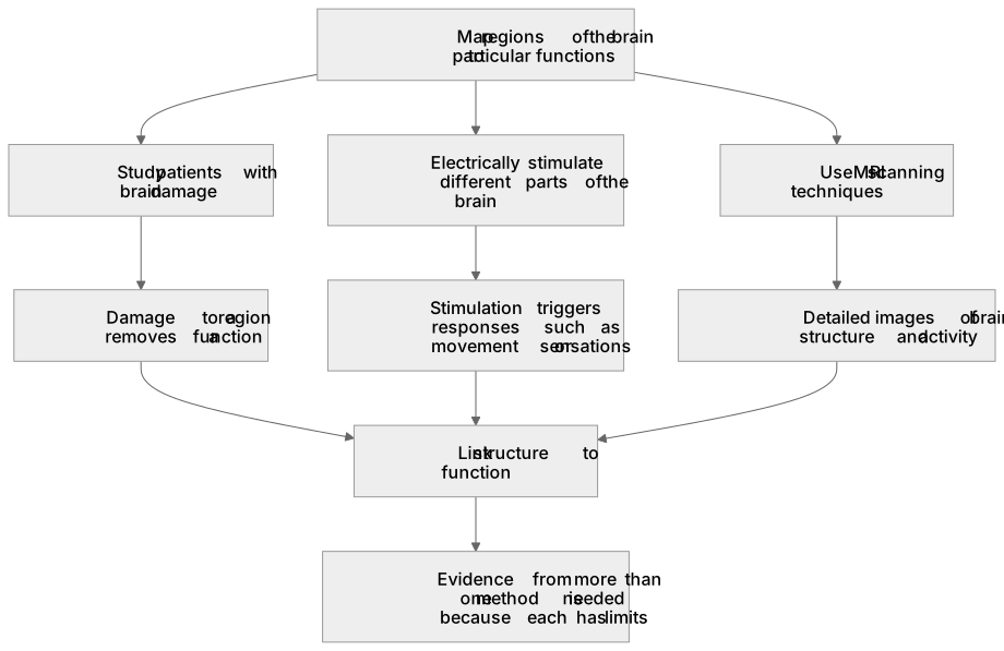
The brain (biology only): (HT only) Neuroscientists have been able to map the regions of the brain to particular functions by studying patients with brain damage, electrically stimulating different parts of the brain and using MRI scanning techniques.
The brain is a complex organ whose regions carry out different functions, but it cannot be studied by simple dissection alone.
Checked against the AQA 8461 specification (opens in a new tab) on .
- Point 4 of 5
- 4 key terms
Study notes
From the checked teaching notes for this specification point.
The idea
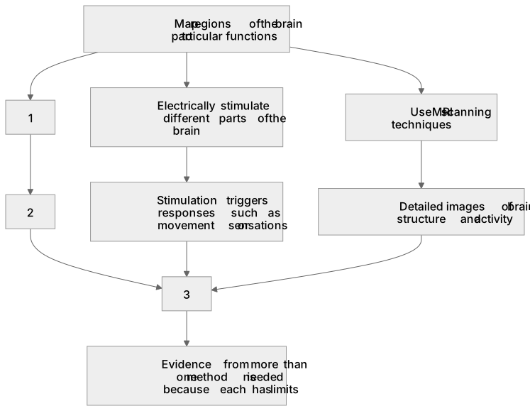
The brain is a complex organ whose regions carry out different functions, but it cannot be studied by simple dissection alone. Neuroscientists combine three approaches. First, studying patients with brain damage: if damage to a region removes a function, that region is likely to control it. For example, damage to the cerebellum may affect balance and coordination. Second, electrically stimulating different parts of the brain during surgery can trigger responses, such as movement or sensations, showing what a region does. Third, MRI scanning techniques produce detailed images of brain structure and activity, allowing living brains to be mapped safely. Together these methods link structure to function, though each has limits.
What earns the marks
- Studying patients with brain damage links a damaged region to a lost function, for example damage to the cerebellum affecting balance and coordination.
- Electrically stimulating different parts of the brain can trigger specific responses, such as movement or a sensation, revealing the function of that region.
- MRI scanning techniques produce detailed images of brain structure and activity, allowing functions to be mapped in living patients.
- Mapping regions to functions requires evidence from more than one method because each method has limitations.
- The three approaches are complementary: damage studies show what is lost, stimulation shows what can be triggered, and MRI shows structure and activity.
- Conclusions are strengthened when findings from different patients and methods agree.
Common mistakes and how to put them right
Thinking that one method alone proves a region's function.
Put it right: Functions are mapped by combining evidence from damage studies, electrical stimulation and MRI scanning.
Confusing MRI with electrical stimulation.
Put it right: MRI scanning produces images of structure and activity, whereas electrical stimulation directly activates brain tissue.
Assuming brain damage always reveals function clearly.
Put it right: Damage may affect several regions or be compensated for, so results must be interpreted alongside other evidence.
Exam tips
- Name all three techniques and give a specific example of what each can reveal.
- Use the phrase 'map regions of the brain to particular functions' to show you understand the overall aim.
- When explaining a limitation, link it to why several methods are used together rather than relying on one.
Try the lesson
The start of the The brain (biology only): (HT only) Neuroscientists have been able to map the regions of the brain to particular functions by studying patients with brain damage, electrically stimulating different parts of the brain and using MRI scanning techniques. lesson, made from checked teaching notes for this specification point.
Have a go first
Neuroscientists can map regions of the brain to particular functions. If a patient loses the ability to understand spoken language after a stroke, which method of brain mapping is being used to link the damaged region to this function?
Listen
The brain is a complex organ whose regions carry out different functions, but it cannot be studied by simple dissection alone. Neuroscientists combine three approaches. First, studying patients with brain damage: if damage to a region removes a function, that region is likely to control it. For example, damage to the cerebellum may affect balance and coordination. Second, electrically stimulating different parts of the brain during surgery can trigger responses, such as movement or sensations, showing what a region does. Third, MRI scanning techniques produce detailed images of brain structure and activity, allowing living brains to be mapped safely. Together these methods link structure to function, though each has limits. A strong answer earns marks for these points. Studying patients with brain damage links a damaged region to a lost function, for example damage to the cerebellum affecting balance and coordination. Electrically stimulating different parts of the brain can trigger specific responses, such as movement or a sensation, revealing the function of that region. MRI scanning techniques produce detailed images of brain structure and activity, allowing functions to be mapped in living patients. Mapping regions to functions requires evidence from more than one method because each method has limitations. A common slip: Thinking that one method alone proves a region's function. Instead, functions are mapped by combining evidence from damage studies, electrical stimulation and MRI scanning. Exam tip: Name all three techniques and give a specific example of what each can reveal.
See it
Key words
Say what each word means before you turn the card.
Studying patients with brain damage
Try answering aloud or in your head before turning the card.
Electrically stimulating different parts of the brain
Try answering aloud or in your head before turning the card.
MRI scanning techniques
Try answering aloud or in your head before turning the card.
Mapping regions to functions
Try answering aloud or in your head before turning the card.
See every definition
- Studying patients with brain damage
- If damage to a region removes a function, that region is likely to control it.
- Electrically stimulating different parts of the brain
- During surgery this can trigger responses, such as movement or sensations, showing what a region does.
- MRI scanning techniques
- These produce detailed images of brain structure and activity, allowing living brains to be mapped safely.
- Mapping regions to functions
- This requires evidence from more than one method because each method has limitations.
Label the diagram
Some boxes have lost their names. Pick the right name for each number.
1Box 1
2Box 2
3Box 3
There is more to this lesson
The full lesson goes on to check your understanding and help you earn the marks. Create a free account to carry on, with your progress saved.
A week of Premium from your first lesson · Then the free plan continues · No card, no charge
Test your understanding of The brain (biology only): (HT only) Neuroscientists have been able to map the regions of the brain to particular functions by studying patients with brain damage, electrically stimulating different parts of the brain and using MRI scanning techniques.
Active recall beats re-reading. Take an instant quiz now to test what you’ve learned and lock it into long-term memory.
A week of Premium from your first lesson · Then the free plan continues · No card, no charge