AQA · GCSE · Biology
Course version: 8461 · Version from Sept 2018
Students should be able to interpret ray diagrams, showing these two…
Full specification wording
The eye (biology only): Students should be able to interpret ray diagrams, showing these two common defects of the eye and demonstrate how spectacle lenses correct them.
Ray diagrams show how light from a point is refracted by the eye and where the image forms.
Checked against the AQA 8461 specification (opens in a new tab) on .
- Point 10 of 10
- 4 key terms
Study notes
From the checked teaching notes for this specification point.
The idea
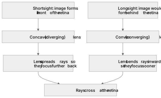
Ray diagrams show how light from a point is refracted by the eye and where the image forms. In short sight (myopia) the image forms in front of the retina, often because the eyeball is too long or the lens is too strong; a concave (diverging) spectacle lens spreads rays before they enter the eye so they focus on the retina. In long sight (hyperopia) the image would form behind the retina, often because the eyeball is too short or the lens too weak; a convex (converging) lens bends rays inward so they focus on the retina. To interpret a diagram, trace rays from the object through the lens, note where they cross, and compare that point with the retina.
What earns the marks
- Short sight: image forms in front of the retina; corrected with a concave (diverging) lens.
- Long sight: image would form behind the retina; corrected with a convex (converging) lens.
- Concave lenses spread rays so they focus further back, onto the retina.
- Convex lenses bend rays inward so they focus sooner, onto the retina.
- Ray diagrams should show rays crossing at the retina after correction.
- Causes include eyeball length and lens strength, not damage to the optic nerve.
Common mistakes and how to put them right
Swapping the lens types.
Put it right: Concave for short sight, convex for long sight.
Drawing corrected rays crossing in front of the retina.
Put it right: They must meet on the retina.
Confusing the defect with its cause.
Put it right: State both the image position and a possible cause.
Exam tips
- Label retina, lens and image position on every diagram.
- Use arrows to show ray direction.
- State the lens type and its effect in one sentence.
Try the lesson
The start of the The eye (biology only): Students should be able to interpret ray diagrams, showing these two common defects of the eye and demonstrate how spectacle lenses correct them. lesson, made from checked teaching notes for this specification point.
Have a go first
A student is interpreting a ray diagram showing an eye defect. The parallel rays of light from a distant object are shown converging to a focal point in front of the retina. Which defect is shown in the diagram, and which type of lens is needed to correct it?
Listen
Ray diagrams show how light from a point is refracted by the eye and where the image forms. In short sight (myopia) the image forms in front of the retina, often because the eyeball is too long or the lens is too strong; a concave (diverging) spectacle lens spreads rays before they enter the eye so they focus on the retina. In long sight (hyperopia) the image would form behind the retina, often because the eyeball is too short or the lens too weak; a convex (converging) lens bends rays inward so they focus on the retina. To interpret a diagram, trace rays from the object through the lens, note where they cross, and compare that point with the retina. A strong answer earns marks for these points. Short sight: image forms in front of the retina; corrected with a concave (diverging) lens. Long sight: image would form behind the retina; corrected with a convex (converging) lens. Concave lenses spread rays so they focus further back, onto the retina. Convex lenses bend rays inward so they focus sooner, onto the retina. A common slip: Swapping the lens types. Instead, concave for short sight, convex for long sight. Exam tip: Label retina, lens and image position on every diagram.
See it
Key words
Say what each word means before you turn the card.
Short sight
Try answering aloud or in your head before turning the card.
Long sight
Try answering aloud or in your head before turning the card.
Concave (diverging) lens
Try answering aloud or in your head before turning the card.
Convex (converging) lens
Try answering aloud or in your head before turning the card.
See every definition
- Short sight
- A defect in which the image forms in front of the retina, often because the eyeball is too long or the lens is too strong.
- Long sight
- A defect in which the image would form behind the retina, often because the eyeball is too short or the lens too weak.
- Concave (diverging) lens
- A spectacle lens that spreads rays before they enter the eye so they focus on the retina, correcting short sight.
- Convex (converging) lens
- A spectacle lens that bends rays inward so they focus on the retina, correcting long sight.
Label the diagram
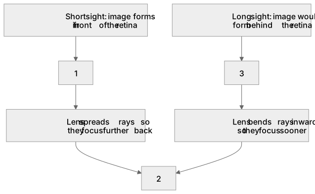
Some boxes have lost their names. Pick the right name for each number.
1Box 1
2Box 2
3Box 3
There is more to this lesson
The full lesson goes on to check your understanding and help you earn the marks. Create a free account to carry on, with your progress saved.
A week of Premium from your first lesson · Then the free plan continues · No card, no charge
Test your understanding of The eye (biology only): Students should be able to interpret ray diagrams, showing these two common defects of the eye and demonstrate how spectacle lenses correct them.
Active recall beats re-reading. Take an instant quiz now to test what you’ve learned and lock it into long-term memory.
A week of Premium from your first lesson · Then the free plan continues · No card, no charge