OCR · A-Level · Biology
Course version: H420 · Version from Sept 2025
(e) (i) the structure and mode of action of phagocytes
Full specification wording
Communicable diseases, disease prevention and the immune system: (e) (i) the structure and mode of action of phagocytes
Phagocytes are white blood cells that engulf and digest pathogens by phagocytosis.
Checked against the OCR H420 specification (opens in a new tab) on .
- Point 5 of 14
- 4 key terms
Study notes
From the checked teaching notes for this specification point.
The idea
Phagocytes are white blood cells that engulf and digest pathogens by phagocytosis. Neutrophils have a lobed nucleus and granular cytoplasm; macrophages are larger, with a rounded or kidney-shaped nucleus, and act as antigen-presenting cells. In phagocytosis the phagocyte is attracted to chemicals from the pathogen, binds it, and the cell-surface membrane invaginates to enclose the microorganism in a vesicle called a phagosome. Lysosomes fuse with the phagosome and release hydrolytic enzymes (lysozymes) that digest the pathogen. Soluble digestion products are absorbed into the cytoplasm. In macrophages, antigens from the digested pathogen are displayed on the cell-surface membrane, helping to activate other immune responses. MCQ items test recognition of these structural features and the correct ordered sequence of phagocytosis, so learn the stages and the organelle roles precisely.
What earns the marks
- Phagocytes are white blood cells of the immune system that carry out phagocytosis.
- Neutrophils have a lobed (multi-lobed) nucleus and granular cytoplasm; macrophages are larger with a rounded or kidney-shaped nucleus.
- The phagocyte is attracted to the pathogen by chemicals it releases and binds to it.
- The cell-surface membrane invaginates (engulfs) the pathogen to form a phagosome vesicle.
- Lysosomes fuse with the phagosome and release hydrolytic enzymes (lysozymes) that digest the pathogen.
- Digested soluble products are absorbed into the phagocyte's cytoplasm.
- Macrophages act as antigen-presenting cells, displaying pathogen antigens on their cell-surface membrane.
Common mistakes and how to put them right
Thinking the pathogen is digested by enzymes in the cytoplasm before being enclosed.
Put it right: Correct that engulfment into a phagosome happens first, then lysosomes fuse and release enzymes.
Confusing phagocytes with lymphocytes.
Put it right: Correct that phagocytes engulf pathogens non-specifically, whereas lymphocytes such as B and T cells give specific responses.
Believing lysosomes are the same as the phagosome.
Put it right: Correct that the phagosome is the vesicle containing the pathogen, and lysosomes are separate organelles that fuse with it.
Assuming all phagocytes present antigens.
Put it right: Correct that macrophages are the main antigen-presenting phagocytes, while neutrophils mainly destroy pathogens.
Exam tips
- Match each structure to its function: lobed nucleus and granules for neutrophils, antigen presentation for macrophages.
- Eliminate options that place digestion before engulfment or that name the wrong organelle.
- Check whether an option describes specific (lymphocyte) or non-specific (phagocyte) immunity before selecting.
Try the lesson
The start of the Communicable diseases, disease prevention and the immune system: (e) (i) the structure and mode of action of phagocytes lesson, made from checked teaching notes for this specification point.
Have a go first
Which sequence correctly describes the mode of action of a phagocyte on a pathogen?
Listen
Phagocytes are white blood cells that engulf and digest pathogens by phagocytosis. Neutrophils have a lobed nucleus and granular cytoplasm; macrophages are larger, with a rounded or kidney-shaped nucleus, and act as antigen-presenting cells. In phagocytosis the phagocyte is attracted to chemicals from the pathogen, binds it, and the cell-surface membrane invaginates to enclose the microorganism in a vesicle called a phagosome. Lysosomes fuse with the phagosome and release hydrolytic enzymes (lysozymes) that digest the pathogen. Soluble digestion products are absorbed into the cytoplasm. In macrophages, antigens from the digested pathogen are displayed on the cell-surface membrane, helping to activate other immune responses. MCQ items test recognition of these structural features and the correct ordered sequence of phagocytosis, so learn the stages and the organelle roles precisely. A strong answer earns marks for these points. Phagocytes are white blood cells of the immune system that carry out phagocytosis. Neutrophils have a lobed (multi-lobed) nucleus and granular cytoplasm; macrophages are larger with a rounded or kidney-shaped nucleus. The phagocyte is attracted to the pathogen by chemicals it releases and binds to it. The cell-surface membrane invaginates (engulfs) the pathogen to form a phagosome vesicle. Exam tip: Match each structure to its function: lobed nucleus and granules for neutrophils, antigen presentation for macrophages.
See it
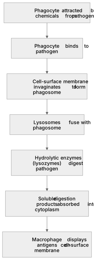
Put it in order
Put these steps in the order they happen.
- 1. Macrophage displays antigens on cell-surface membrane
- 2. Soluble digestion products absorbed into cytoplasm
- 3. Cell-surface membrane invaginates to form phagosome
- 4. Hydrolytic enzymes (lysozymes) digest pathogen
- 5. Phagocyte attracted by chemicals from pathogen
- 6. Phagocyte binds to pathogen
- 7. Lysosomes fuse with phagosome
Key words
Say what each word means before you turn the card.
Phagocytosis
Try answering aloud or in your head before turning the card.
Phagosome
Try answering aloud or in your head before turning the card.
Lysosomes
Try answering aloud or in your head before turning the card.
Antigen-presenting cells
Try answering aloud or in your head before turning the card.
See every definition
- Phagocytosis
- The process by which phagocytes engulf and digest pathogens, with the cell-surface membrane invaginating to enclose the microorganism in a phagosome.
- Phagosome
- A vesicle formed when the cell-surface membrane invaginates to enclose the microorganism; lysosomes fuse with it and release hydrolytic enzymes.
- Lysosomes
- Organelles that fuse with the phagosome and release hydrolytic enzymes (lysozymes) that digest the pathogen.
- Antigen-presenting cells
- Macrophages display pathogen antigens from the digested pathogen on their cell-surface membrane, helping to activate other immune responses.
Label the diagram
Some boxes have lost their names. Pick the right name for each number.
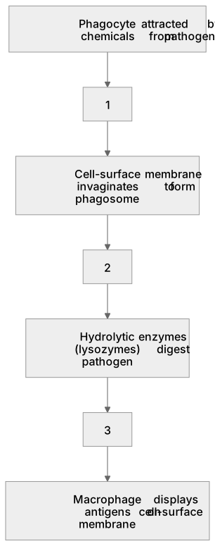
1Box 1
2Box 2
3Box 3
There is more to this lesson
The full lesson goes on to check your understanding and help you earn the marks. Create a free account to carry on, with your progress saved.
A week of Premium from your first lesson · Then the free plan continues · No card, no charge
Test your understanding of Communicable diseases, disease prevention and the immune system: (e) (i) the structure and mode of action of phagocytes
Active recall beats re-reading. Take an instant quiz now to test what you’ve learned and lock it into long-term memory.
A week of Premium from your first lesson · Then the free plan continues · No card, no charge