OCR · A-Level · Biology
Course version: H420 · Version from Sept 2025
(b) (i) the structure and functions of the mammalian liver
Full specification wording
Excretion as an example of homeostatic control: (b) (i) the structure and functions of the mammalian liver
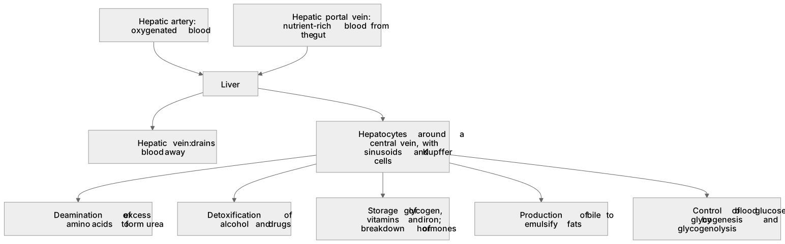
The liver is a large lobed organ supplied by the hepatic artery (oxygenated blood) and hepatic portal vein (nutrient-rich blood from the gut), drained by the hepatic vein.
Checked against the OCR H420 specification (opens in a new tab) on .
- Point 2 of 6
- 3 key terms
Study notes
From the checked teaching notes for this specification point.
The idea
The liver is a large lobed organ supplied by the hepatic artery (oxygenated blood) and hepatic portal vein (nutrient-rich blood from the gut), drained by the hepatic vein. It is built from hepatocytes arranged around a central vein, with sinusoids and Kupffer cells. Its functions include: formation of urea from excess amino acids by deamination; detoxification of substances such as alcohol and drugs; storage of glycogen, vitamins and iron; breakdown of hormones; production of bile for fat emulsification; and control of blood glucose by glycogenesis and glycogenolysis. A method is to link each function to the structure that performs it, for example hepatocytes for deamination and glycogen storage.
What earns the marks
- Blood supply: hepatic artery brings oxygenated blood, hepatic portal vein brings nutrients from the gut, hepatic vein drains blood.
- Structure: hepatocytes arranged around a central vein, with sinusoids and Kupffer cells.
- Deamination of excess amino acids to form urea, which is passed to the kidneys.
- Detoxification of alcohol and drugs.
- Storage of glycogen, vitamins and iron; breakdown of hormones.
- Production of bile to emulsify fats; role in controlling blood glucose by glycogenesis and glycogenolysis.
Common mistakes and how to put them right
Confusing the hepatic portal vein with the hepatic vein.
Put it right: The portal vein brings blood to the liver, the hepatic vein drains it away.
Thinking the liver makes urea from ammonia directly without deamination.
Put it right: Deamination of amino acids produces ammonia, which is converted to urea.
Assuming the liver only stores glycogen.
Put it right: It also detoxifies, deaminates, stores vitamins and iron, and produces bile.
Exam tips
- Link each function to a named structure or blood vessel.
- Use the terms deamination, detoxification, glycogenesis and glycogenolysis precisely.
- Remember the liver is part of excretion because it forms urea, even though the kidneys excrete it.
Try the lesson
The start of the Excretion as an example of homeostatic control: (b) (i) the structure and functions of the mammalian liver lesson, made from checked teaching notes for this specification point.
Have a go first
Which blood vessel carries oxygenated blood from the heart directly to the liver?
Listen
The liver is a large lobed organ supplied by the hepatic artery (oxygenated blood) and hepatic portal vein (nutrient-rich blood from the gut), drained by the hepatic vein. It is built from hepatocytes arranged around a central vein, with sinusoids and Kupffer cells. Its functions include: formation of urea from excess amino acids by deamination; detoxification of substances such as alcohol and drugs; storage of glycogen, vitamins and iron; breakdown of hormones; production of bile for fat emulsification; and control of blood glucose by glycogenesis and glycogenolysis. A method is to link each function to the structure that performs it, for example hepatocytes for deamination and glycogen storage. A strong answer earns marks for these points. Blood supply: hepatic artery brings oxygenated blood, hepatic portal vein brings nutrients from the gut, hepatic vein drains blood. Structure: hepatocytes arranged around a central vein, with sinusoids and Kupffer cells. Deamination of excess amino acids to form urea, which is passed to the kidneys. Detoxification of alcohol and drugs. A common slip: Confusing the hepatic portal vein with the hepatic vein. Instead, the portal vein brings blood to the liver, the hepatic vein drains it away. Exam tip: Link each function to a named structure or blood vessel.
See it
Key words
Say what each word means before you turn the card.
Hepatic portal vein
Try answering aloud or in your head before turning the card.
Hepatocytes
Try answering aloud or in your head before turning the card.
Glycogenesis and glycogenolysis
Try answering aloud or in your head before turning the card.
See every definition
- Hepatic portal vein
- Vessel that brings nutrient-rich blood from the gut to the liver.
- Hepatocytes
- Cells arranged around a central vein, which perform deamination and glycogen storage.
- Glycogenesis and glycogenolysis
- Processes by which the liver controls blood glucose.
Label the diagram
Some boxes have lost their names. Pick the right name for each number.
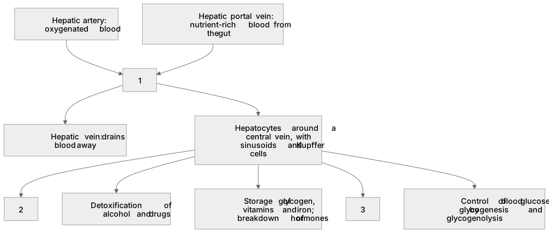
1Box 1
2Box 2
3Box 3
There is more to this lesson
The full lesson goes on to check your understanding and help you earn the marks. Create a free account to carry on, with your progress saved.
A week of Premium from your first lesson · Then the free plan continues · No card, no charge
Test your understanding of Excretion as an example of homeostatic control: (b) (i) the structure and functions of the mammalian liver
Active recall beats re-reading. Take an instant quiz now to test what you’ve learned and lock it into long-term memory.
A week of Premium from your first lesson · Then the free plan continues · No card, no charge