OCR · A-Level · Physics
Course version: H556 · Version from Sept 2015
(b) gamma camera; components – collimator, scintillator, photomultiplier…
Full specification wording
Diagnostic methods in medicine: (b) gamma camera; components – collimator, scintillator, photomultiplier tubes, computer and display; formation of image
A gamma camera images a patient after a gamma-emitting tracer is administered.
Checked against the OCR H556 specification (opens in a new tab) on .
- Point 2 of 5
- 4 key terms
Study notes
From the checked teaching notes for this specification point.
The idea
A gamma camera images a patient after a gamma-emitting tracer is administered. The collimator is a lead block with parallel holes that admits only near-parallel gamma rays, so each point on the detector maps to a known direction. Gamma photons strike a large scintillator crystal, commonly sodium iodide doped with thallium, producing many visible-light photons. These reach an array of photomultiplier tubes; each tube's photocathode releases electrons by the photoelectric effect, and dynodes multiply the signal by secondary emission. The computer compares pulse heights across tubes to calculate the interaction position, building a two-dimensional image of tracer distribution. The display shows counts as brightness or colour, so regions of high uptake appear bright. The collimator limits sensitivity but preserves spatial resolution.
What earns the marks
- The collimator is a lead grid with parallel channels that absorbs oblique gamma rays so only near-parallel rays reach the scintillator, defining the line of sight.
- The scintillator, often NaI(Tl), absorbs a gamma photon and emits a burst of visible-light photons whose number is proportional to the deposited energy.
- Photomultiplier tubes convert light into an electrical pulse: the photocathode emits electrons, and dynodes produce secondary emission, multiplying the signal.
- The computer uses the relative pulse sizes from several photomultiplier tubes to locate each scintillation event and accumulates counts to form the image.
- The display presents the count distribution as brightness or colour, allowing regions of high and low tracer uptake to be distinguished.
Common mistakes and how to put them right
Thinking the collimator focuses gamma rays like a lens.
Put it right: It does not refract or focus, it absorbs oblique rays so only near-parallel rays pass.
Confusing the scintillator with the photomultiplier tube.
Put it right: The scintillator converts gamma photons to visible light, while the photomultiplier tube converts light to an electrical pulse.
Believing the gamma camera detects positrons or emits radiation.
Put it right: It passively detects gamma photons emitted from within the patient.
Assuming the computer produces a three-dimensional image.
Put it right: A standard gamma camera produces a two-dimensional projection image.
Exam tips
- Link each component to its function in one sentence: collimator selects direction, scintillator converts gamma to light, photomultiplier tubes amplify and convert to electrical signal, computer locates events, display shows the image.
- Use the correct order of energy conversion: gamma photon to visible photons to electrons to electrical pulse.
- When explaining image formation, state that the computer uses the relative signals from several photomultiplier tubes to calculate the position of each event.
Try the lesson
The start of the Diagnostic methods in medicine: (b) gamma camera; components – collimator, scintillator, photomultiplier tubes, computer and display; formation of image lesson, made from checked teaching notes for this specification point.
Listen
A gamma camera images a patient after a gamma-emitting tracer is administered. The collimator is a lead block with parallel holes that admits only near-parallel gamma rays, so each point on the detector maps to a known direction. Gamma photons strike a large scintillator crystal, commonly sodium iodide doped with thallium, producing many visible-light photons. These reach an array of photomultiplier tubes; each tube's photocathode releases electrons by the photoelectric effect, and dynodes multiply the signal by secondary emission. The computer compares pulse heights across tubes to calculate the interaction position, building a two-dimensional image of tracer distribution. The display shows counts as brightness or colour, so regions of high uptake appear bright. The collimator limits sensitivity but preserves spatial resolution. A strong answer earns marks for these points. The collimator is a lead grid with parallel channels that absorbs oblique gamma rays so only near-parallel rays reach the scintillator, defining the line of sight. The scintillator, often NaI(Tl), absorbs a gamma photon and emits a burst of visible-light photons whose number is proportional to the deposited energy. Photomultiplier tubes convert light into an electrical pulse: the photocathode emits electrons, and dynodes produce secondary emission, multiplying the signal. The computer uses the relative pulse sizes from several photomultiplier tubes to locate each scintillation event and accumulates counts to form the image. Exam tip: Link each component to its function in one sentence: collimator selects direction, scintillator converts gamma to light, photomultiplier tubes amplify and convert to electrical signal, computer locates events, display shows the image.
See it
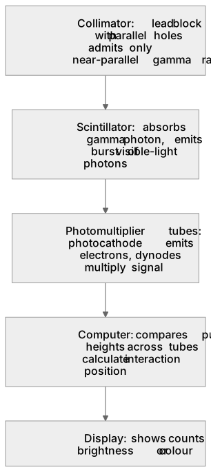
Put it in order
Put these steps in the order they happen.
- 1. Scintillator: absorbs gamma photon, emits burst of visible-light photons
- 2. Photomultiplier tubes: photocathode emits electrons, dynodes multiply signal
- 3. Collimator: lead block with parallel holes admits only near-parallel gamma rays
- 4. Display: shows counts as brightness or colour
- 5. Computer: compares pulse heights across tubes to calculate interaction position
Key words
Say what each word means before you turn the card.
Collimator
Try answering aloud or in your head before turning the card.
Scintillator
Try answering aloud or in your head before turning the card.
Photomultiplier tubes
Try answering aloud or in your head before turning the card.
Computer
Try answering aloud or in your head before turning the card.
See every definition
- Collimator
- A lead block with parallel holes that admits only near-parallel gamma rays, so each point on the detector maps to a known direction.
- Scintillator
- A large crystal, commonly sodium iodide doped with thallium, that gamma photons strike to produce many visible-light photons.
- Photomultiplier tubes
- An array of tubes in which each tube's photocathode releases electrons by the photoelectric effect and dynodes multiply the signal by secondary emission.
- Computer
- Compares pulse heights across tubes to calculate the interaction position, building a two-dimensional image of tracer distribution.
Label the diagram
Some boxes have lost their names. Pick the right name for each number.
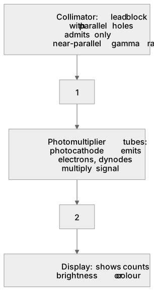
1Box 1
2Box 2
There is more to this lesson
The full lesson goes on to check your understanding and help you earn the marks. Create a free account to carry on, with your progress saved.
A week of Premium from your first lesson · Then the free plan continues · No card, no charge
Test your understanding of Diagnostic methods in medicine: (b) gamma camera; components – collimator, scintillator, photomultiplier tubes, computer and display; formation of image
Active recall beats re-reading. Take an instant quiz now to test what you’ve learned and lock it into long-term memory.
A week of Premium from your first lesson · Then the free plan continues · No card, no charge