OCR · A-Level · Chemistry
Course version: H432 · Version from Sept 2017
Spectroscopy: (ii) mass spectra (see also 4.2.4 f–g)
Mass spectrometry ionises a sample, accelerates the ions and separates them by mass-to-charge ratio, m/z.
- Point 14 of 16
- 4 key terms
Study notes
From the teaching notes for this specification point.
The idea
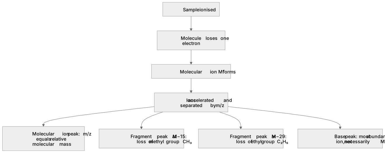
Mass spectrometry ionises a sample, accelerates the ions and separates them by mass-to-charge ratio, m/z. In organic chemistry the key skill is reading a mass spectrum to find the molecular ion, M⁺, which gives the relative molecular mass, and to interpret fragmentation peaks that reveal structural features. The molecular ion forms when a molecule loses one electron, so its m/z equals the relative molecular mass for a singly charged ion. A peak at M−15 often signals loss of a methyl group, CH₃, and M−29 can indicate loss of an ethyl group, C₂H₅. The base peak is simply the most abundant ion, not necessarily M⁺. This links back to 4.2.4 f–g, where you met ionisation and the mass spectrometer, so apply that instrumental knowledge here to deduce structure.
What earns the marks
- Identifies the molecular ion peak as the peak whose m/z value equals the relative molecular mass of the compound.
- Explains that the molecular ion forms when a molecule loses one electron to give a singly charged positive ion.
- Uses a fragment peak at M−15 to suggest loss of a methyl group from the molecular ion.
- Uses a fragment peak at M−29 to suggest loss of an ethyl group from the molecular ion.
- Distinguishes the base peak as the most abundant ion rather than assuming it is the molecular ion.
- Links the ionisation and separation steps to the earlier study of the mass spectrometer.
Common mistakes and how to put them right
Assumes the tallest peak is always the molecular ion.
Put it right: Check the highest m/z peak for M⁺ and treat the tallest as the base peak.
Ignores the charge when reading m/z.
Put it right: m/z is mass-to-charge ratio, so a singly charged ion gives m/z equal to its mass.
Treats every fragment as random.
Put it right: Look for characteristic losses such as M−15 and M−29 to infer groups present.
Exam tips
- Locate the molecular ion first by finding the peak at highest m/z that is consistent with the molecule, then work down through fragments.
- Quote the m/z value and state the group lost, for example a peak 15 units below M⁺ suggests loss of CH₃.
- Check whether the question asks for molecular mass, a fragment or structural evidence, and answer that specific point.
Try the lesson
The start of the Spectroscopy: (ii) mass spectra (see also 4.2.4 f–g) lesson, made from teaching notes for this specification point.
Listen
Mass spectrometry ionises a sample, accelerates the ions and separates them by mass-to-charge ratio, m/z. In organic chemistry the key skill is reading a mass spectrum to find the molecular ion, M⁺, which gives the relative molecular mass, and to interpret fragmentation peaks that reveal structural features. The molecular ion forms when a molecule loses one electron, so its m/z equals the relative molecular mass for a singly charged ion. A peak at M−15 often signals loss of a methyl group, CH₃, and M−29 can indicate loss of an ethyl group, C₂H₅. The base peak is simply the most abundant ion, not necessarily M⁺. This links back to 4.2.4 f–g, where you met ionisation and the mass spectrometer, so apply that instrumental knowledge here to deduce structure. A strong answer earns marks for these points. Identifies the molecular ion peak as the peak whose m/z value equals the relative molecular mass of the compound. Explains that the molecular ion forms when a molecule loses one electron to give a singly charged positive ion. Uses a fragment peak at M−15 to suggest loss of a methyl group from the molecular ion. Uses a fragment peak at M−29 to suggest loss of an ethyl group from the molecular ion. Exam tip: Locate the molecular ion first by finding the peak at highest m/z that is consistent with the molecule, then work down through fragments.
See it
Key words
Say what each word means before you turn the card.
Molecular ion
Try answering aloud or in your head before turning the card.
Base peak
Try answering aloud or in your head before turning the card.
Fragmentation
Try answering aloud or in your head before turning the card.
Ionisation and separation
Try answering aloud or in your head before turning the card.
See every definition
- Molecular ion
- Forms when a molecule loses one electron, so its m/z equals the relative molecular mass for a singly charged ion.
- Base peak
- The most abundant ion, which is not necessarily the molecular ion.
- Fragmentation
- Fragment peaks such as M−15 and M−29 reveal structural features, suggesting loss of a methyl group or an ethyl group.
- Ionisation and separation
- The sample is ionised, the ions are accelerated and separated by mass-to-charge ratio, m/z.
Label the diagram
Some boxes have lost their names. Pick the right name for each number.
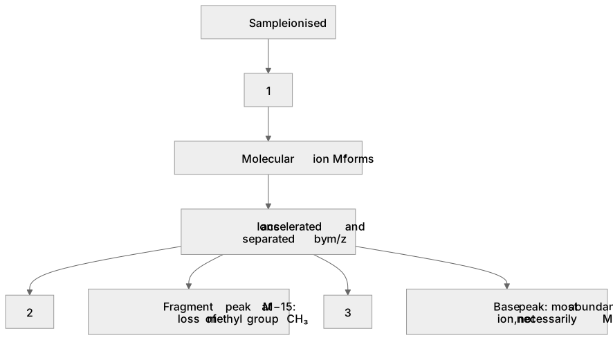
1Box 1
2Box 2
3Box 3
There is more to this lesson
The full lesson goes on to check your understanding and help you earn the marks. Create a free account to carry on, with your progress saved.
A week of Premium from your first lesson · Then the free plan continues · No card, no charge
Test your understanding of Spectroscopy: (ii) mass spectra (see also 4.2.4 f–g)
Active recall beats re-reading. Take an instant quiz now to test what you’ve learned and lock it into long-term memory.
A week of Premium from your first lesson · Then the free plan continues · No card, no charge