Cell level systems — OCR GCSE Study Guide
Exam Board: OCR | Level: GCSE
This guide provides a comprehensive overview of OCR GCSE Combined Science Topic B1: Cell Level Systems. It covers the fundamental differences between cell types, the mechanisms of enzymes, and the core bioenergetic processes of respiration and photosynthesis, all tailored to maximise your exam performance.

## Overview
Welcome to the fascinating world of Cell Level Systems! This topic, B1 in your OCR Gateway specification, is the bedrock of modern biology. It explores the microscopic universe within all living organisms, from the simplest bacteria to complex beings like us. We will delve into the structure of different cell types, understanding how their specialised forms are perfectly adapted for their functions. A major focus for examiners is your ability to link the presence of specific organelles, like mitochondria or chloroplasts, to the vital metabolic processes they perform, such as respiration and photosynthesis. This topic also introduces the elegant lock-and-key mechanism of enzyme action and the factors that can affect it. Success here requires not just memorising facts (AO1), but also applying your knowledge to interpret data, perform calculations, and analyse experimental results (AO2 and AO3). Mastering these foundational concepts is essential, as they provide the synoptic links to many other areas of your GCSE course, including genetics, ecology, and health.
{{asset:cell_level_systems_podcast.mp3}}
## Key Concepts
### Concept 1: Prokaryotic vs. Eukaryotic Cells
The living world is fundamentally divided into two types of cells: prokaryotic and eukaryotic. This distinction is a cornerstone of biology and a frequent subject of exam questions. Credit is consistently given for clearly identifying the key differences.
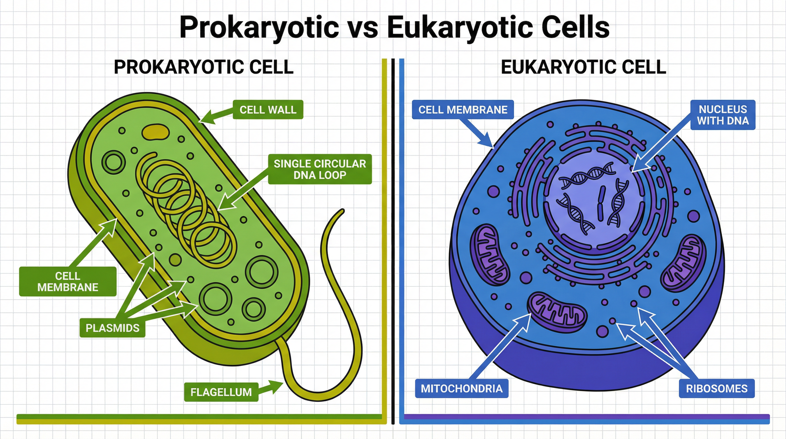
**Prokaryotic Cells**: These are simpler, more ancient cells, with bacteria being the primary example. Their defining feature is the **absence of a true nucleus**. Instead, their genetic material consists of a **single circular DNA loop** that floats freely in the cytoplasm. They may also contain smaller rings of DNA called **plasmids**, which often carry genes for antibiotic resistance. They possess a cell membrane, cytoplasm, and ribosomes, and are surrounded by a cell wall (made of peptidoglycan, not cellulose).
**Eukaryotic Cells**: These are larger, more complex cells that make up all animals, plants, fungi, and protists. Their hallmark is the presence of a **membrane-bound nucleus**, which houses the cell's DNA in the form of linear chromosomes. Eukaryotic cells are highly organised, containing numerous organelles that perform specific functions. For your exam, you must be able to identify and state the function of the nucleus, cytoplasm, cell membrane, mitochondria, and ribosomes in animal cells. For plant cells, you must also know the cell wall, permanent vacuole, and chloroplasts.
### Concept 2: Microscopy and Magnification
Microscopy is the tool that allows us to study cells. For your exam, you need to understand the principles of light microscopy and be able to perform calculations related to magnification. This is a key area where AO2 (application) skills are tested.
**Magnification vs. Resolution**: It is crucial not to confuse these two terms. **Magnification** is how much larger an image appears compared to the actual object. **Resolution** is the ability to distinguish between two separate points; it determines the level of detail that can be seen. While a light microscope can magnify objects up to 1500x, its resolution is limited. Electron microscopes offer much higher magnification and resolution, allowing us to see subcellular structures in fine detail.
**The Magnification Formula**: This is a formula you **must memorise** and be confident in using and rearranging. It is a frequent source of calculation marks.
`Image Size = Actual Size × Magnification`
Or, more simply: `I = A × M`
Examiners will often ask you to calculate one of these values when given the other two. The most common pitfall is failing to ensure units are consistent. You MUST convert all measurements to the same unit before performing the calculation. The standard unit for cell measurements is the micrometre (µm).
**Unit Conversions (Must Memorise)**:
- 1 millimetre (mm) = 1000 micrometres (µm)
- 1 micrometre (µm) = 1000 nanometres (nm)
To convert from mm to µm, you multiply by 1000. To convert from µm to mm, you divide by 1000.
### Concept 3: Enzyme Action
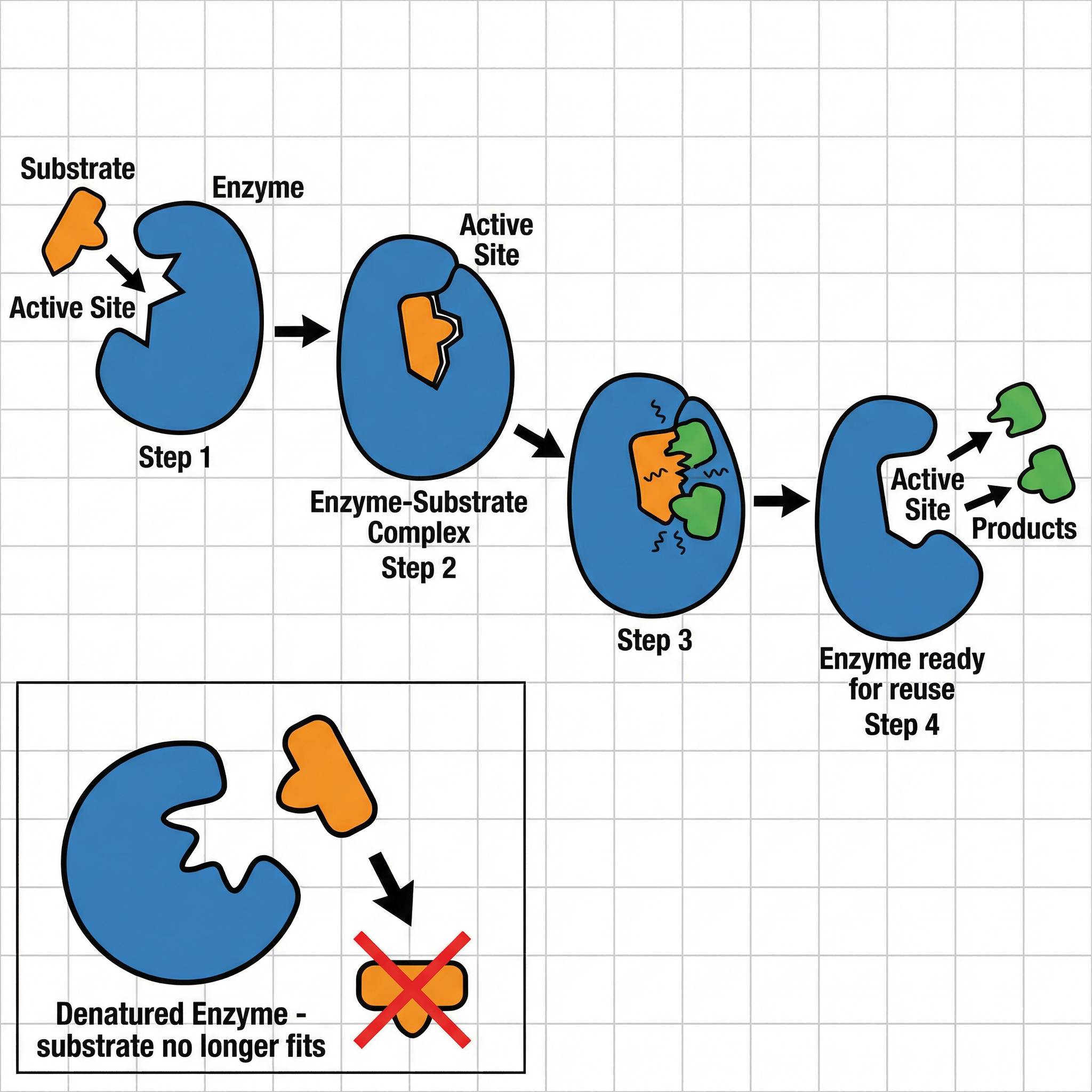
Enzymes are biological catalysts – proteins that speed up metabolic reactions without being used up themselves. Their function is critical to life, and understanding how they work is essential for your exam.
**The Lock-and-Key Model**: This model explains the specificity of enzymes. Each enzyme has a uniquely shaped **active site**. The **substrate** (the molecule the enzyme acts on) has a complementary shape. The substrate fits into the active site, much like a key fits into a lock, forming an **enzyme-substrate complex**. The reaction then occurs, and the products are released. The enzyme is unchanged and can be used again.
**Factors Affecting Enzyme Activity**: Examiners frequently test your understanding of how temperature and pH affect enzyme function, often through graph interpretation questions.
- **Temperature**: As temperature increases, enzyme activity increases because molecules have more kinetic energy, leading to more frequent collisions between the enzyme and substrate. This continues until the **optimum temperature** is reached. Beyond this point, the enzyme starts to **denature**. The high temperature breaks the bonds holding the protein in its specific 3D shape. The active site changes shape permanently, and the substrate can no longer fit. It is vital to use the term 'denatured', not 'killed' or 'died'.
- **pH**: Each enzyme has an **optimum pH** at which it works best. For example, pepsin in the acidic environment of the stomach has an optimum pH of 2. If the pH is too high or too low, it interferes with the bonds in the enzyme, causing it to denature and its active site to change shape.
### Concept 4: Bioenergetics - Photosynthesis and Respiration
This section covers how organisms obtain and use energy. These processes are fundamental to life and are heavily tested.
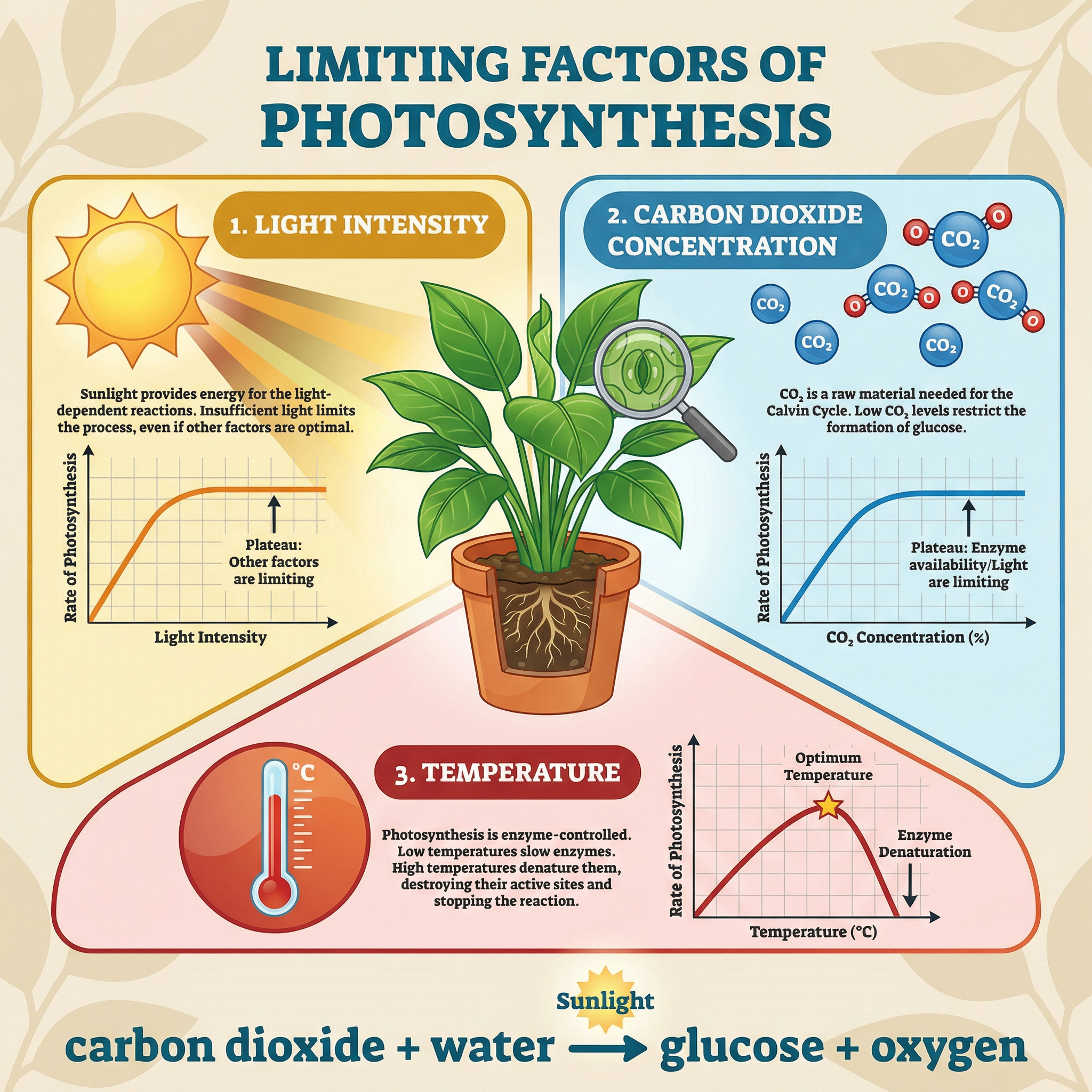
**Photosynthesis**: This is the process where plants and algae use light energy to convert carbon dioxide and water into glucose and oxygen. It occurs in the **chloroplasts**.
- **Word Equation (Must Memorise)**: `Carbon dioxide + Water → Glucose + Oxygen` (in the presence of light)
- **Symbol Equation (Must Memorise)**: `6CO₂ + 6H₂O → C₆H₁₂O₆ + 6O₂`
**Limiting Factors of Photosynthesis**: The rate of photosynthesis can be limited by light intensity, carbon dioxide concentration, or temperature. In a 6-mark question, you would be expected to explain how each of these factors affects the rate and why it can be described as 'limiting'. For example, on a bright, warm day, the amount of CO₂ in the air might be the limiting factor.
**Aerobic Respiration**: This is the process that releases energy from glucose in the presence of oxygen. It occurs primarily in the **mitochondria**. This is why cells with high energy requirements (e.g., muscle cells, sperm cells) have a large number of mitochondria – a classic structure-function link that earns marks.
- **Word Equation (Must Memorise)**: `Glucose + Oxygen → Carbon dioxide + Water` (+ energy released)
- **Symbol Equation (Must Memorise)**: `C₆H₁₂O₆ + 6O₂ → 6CO₂ + 6H₂O`
Crucially, respiration **releases** energy; it does not 'create' or 'make' energy. This is a fine point of terminology that distinguishes top candidates.
## Mathematical/Scientific Relationships
- **Magnification Formula**: `Image Size = Actual Size × Magnification` (I = A × M). This is not given on the formula sheet and **must be memorised**.
- **Photosynthesis Word Equation**: `Carbon dioxide + Water → Glucose + Oxygen`. **Must be memorised**.
- **Photosynthesis Symbol Equation**: `6CO₂ + 6H₂O → C₆H₁₂O₆ + 6O₂`. **Must be memorised**.
- **Aerobic Respiration Word Equation**: `Glucose + Oxygen → Carbon dioxide + Water`. **Must be memorised**.
- **Aerobic Respiration Symbol Equation**: `C₆H₁₂O₆ + 6O₂ → 6CO₂ + 6H₂O`. **Must be memorised**.
## Practical Applications
This topic includes a required practical investigating the effect of a factor (e.g., light intensity) on the rate of photosynthesis, often using an aquatic plant like Elodea (pondweed).
- **Apparatus**: Pondweed, beaker of water, light source (lamp), ruler, stopwatch.
- **Method**: The rate of photosynthesis is measured by counting the number of oxygen bubbles produced per minute. The lamp is placed at a specific distance (e.g., 10 cm) from the beaker, and the bubbles are counted for one minute. This is repeated to get a mean. The distance is then changed (e.g., 20 cm, 30 cm), and the process is repeated. The light intensity is related to the distance (Intensity ∝ 1/distance²).
- **Common Errors**: Not waiting for the plant to acclimatise to the new light intensity before starting to count; miscounting bubbles; the temperature of the water changing due to the heat from the lamp (a heat shield can be used to prevent this).
- **How it's Tested**: Examiners may ask you to describe the method, identify variables (independent, dependent, control), plot the results on a graph, and draw conclusions. They may also ask you to evaluate the method and suggest improvements.