OCR · A-Level · Physics
Course version: H556 · Version from Sept 2015
Using ultrasound: (c) ultrasound A-scan and B-scan
Ultrasound A-scan and B-scan are two display modes used in medical imaging.
Checked against the OCR H556 specification (opens in a new tab) on .
- Point 3 of 8
- 4 key terms
Study notes
From the checked teaching notes for this specification point.
The idea
Ultrasound A-scan and B-scan are two display modes used in medical imaging. An A-scan (amplitude scan) displays the amplitude of reflected ultrasound pulses against time or depth on a single axis, producing a one-dimensional trace with peaks corresponding to boundaries between media. It is used, for example, in measuring the length of the eye or detecting flaws in materials. A B-scan (brightness scan) uses the same pulse-echo principle but displays the intensity of reflections as brightness on a two-dimensional image, building up a cross-sectional picture by scanning the transducer across the region. Remember that A-scan gives depth information along a line, while B-scan gives a 2D image.
What earns the marks
- A-scan displays reflected pulse amplitude against time or depth on a one-dimensional trace.
- B-scan displays reflection intensity as brightness to form a two-dimensional image.
- A-scan is used for depth measurement along a single line, such as eye length or flaw detection.
- B-scan is used for cross-sectional imaging, such as foetal scanning.
Common mistakes and how to put them right
Confusing A-scan and B-scan displays.
Put it right by remembering that A-scan shows amplitude versus depth (1D) and B-scan shows brightness (2D).
Thinking B-scan does not use pulses.
Put it right by stating that both modes use pulsed ultrasound and detect reflections.
Assuming A-scan produces a two-dimensional image.
Put it right by noting that A-scan is a one-dimensional trace of amplitude against time or depth.
Exam tips
- Use the first letter as a memory aid: A for amplitude, B for brightness.
- Link each scan to a typical application: A-scan for distance measurement, B-scan for imaging.
- Check whether the question asks about the display method or the underlying principle; both use pulse-echo.
Try the lesson
The start of the Using ultrasound: (c) ultrasound A-scan and B-scan lesson, made from checked teaching notes for this specification point.
Listen
Ultrasound A-scan and B-scan are two display modes used in medical imaging. An A-scan (amplitude scan) displays the amplitude of reflected ultrasound pulses against time or depth on a single axis, producing a one-dimensional trace with peaks corresponding to boundaries between media. It is used, for example, in measuring the length of the eye or detecting flaws in materials. A B-scan (brightness scan) uses the same pulse-echo principle but displays the intensity of reflections as brightness on a two-dimensional image, building up a cross-sectional picture by scanning the transducer across the region. Remember that A-scan gives depth information along a line, while B-scan gives a 2D image. A strong answer earns marks for these points. A-scan displays reflected pulse amplitude against time or depth on a one-dimensional trace. B-scan displays reflection intensity as brightness to form a two-dimensional image. A-scan is used for depth measurement along a single line, such as eye length or flaw detection. B-scan is used for cross-sectional imaging, such as foetal scanning. A common slip: Confusing A-scan and B-scan displays. Instead, remembering that A-scan shows amplitude versus depth (1D) and B-scan shows brightness (2D). Exam tip: Use the first letter as a memory aid: A for amplitude, B for brightness.
See it
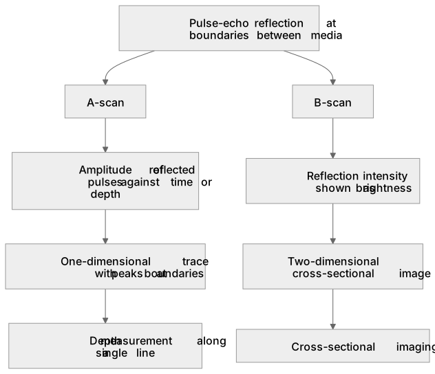
Key words
Say what each word means before you turn the card.
A-scan
Try answering aloud or in your head before turning the card.
B-scan
Try answering aloud or in your head before turning the card.
Pulse-echo principle
Try answering aloud or in your head before turning the card.
Medical imaging
Try answering aloud or in your head before turning the card.
See every definition
- A-scan
- Displays the amplitude of reflected ultrasound pulses against time or depth on a single axis, producing a one-dimensional trace with peaks corresponding to boundaries between media.
- B-scan
- Displays the intensity of reflections as brightness on a two-dimensional image, building up a cross-sectional picture by scanning the transducer across the region.
- Pulse-echo principle
- The principle, used by both A-scan and B-scan, of detecting reflections of pulsed ultrasound at boundaries between media.
- Medical imaging
- The use of display modes such as A-scan and B-scan, for example in measuring the length of the eye or foetal scanning.
Label the diagram
Some boxes have lost their names. Pick the right name for each number.
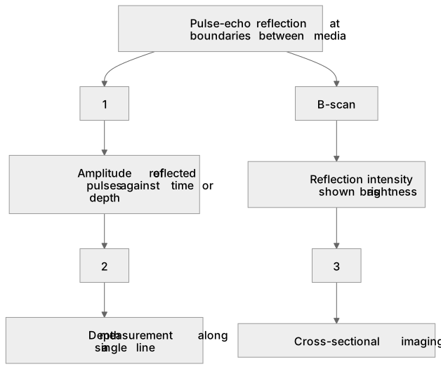
1Box 1
2Box 2
3Box 3
There is more to this lesson
The full lesson goes on to check your understanding and help you earn the marks. Create a free account to carry on, with your progress saved.
A week of Premium from your first lesson · Then the free plan continues · No card, no charge
Test your understanding of Using ultrasound: (c) ultrasound A-scan and B-scan
Active recall beats re-reading. Take an instant quiz now to test what you’ve learned and lock it into long-term memory.
A week of Premium from your first lesson · Then the free plan continues · No card, no charge