OCR · GCSE · Biology
Course version: J247 · Version from Sept 2018
Explain how the main structures of the eye are related to their functions…
Full specification wording
Coordination and control – the nervous system: explain how the main structures of the eye are related to their functions (to include: cornea, iris, pupil, lens, retina, optic nerve, ciliary body, suspensory ligaments) (content found only in the biology separate science qualification)
The mammalian eye is a sensory organ specialised for photoreception and image formation.
- Point 5 of 9
- 3 key terms
Study notes
From the teaching notes for this specification point.
The idea
The mammalian eye is a sensory organ specialised for photoreception and image formation. Light enters through the transparent cornea, which provides the primary refraction. The coloured iris contains circular and radial antagonistic smooth muscles that alter pupil diameter to regulate light entry during the pupillary reflex. The biconvex, flexible crystalline lens provides variable refraction to focus light sharply onto the retina. The retina contains light-sensitive rods for vision in dim illumination and cones for color visual acuity. The ciliary body contains circular ciliary muscles connected to the lens by suspensory ligaments; these control lens curvature during accommodation. The optic nerve conducts sensory impulses to the brain.
What earns the marks
- Describe the cornea as a transparent curved layer that refracts light entering the eye.
- Explain how antagonistic contraction of circular and radial iris muscles regulates pupil diameter in bright versus dim light.
- Describe accommodation for near vision: ciliary muscles contract, suspensory ligaments slacken, and the elastic lens becomes more convex.
- State that the retina contains photoreceptor rods and cones that convert light energy into electrical impulses carried by the optic nerve.
Common mistakes and how to put them right
Believing the lens does most of the light refraction, whereas the cornea refracts the greatest proportion of light.
Stating that suspensory ligaments contract or relax, when they are non-muscular tissues that only tighten or slacken.
Confusing the roles of rods and cones in visual perception.
Exam tips
- Use a memory aid for accommodation: Ciliary muscles Contract, Suspensory ligaments Slacken, and the Lens becomes Rounded and thicker for near objects.
- Never state that ligaments contract; muscles contract, while ligaments merely tighten under tension or slacken.
Try the lesson
The start of the Coordination and control – the nervous system: explain how the main structures of the eye are related to their functions (to include: cornea, iris, pupil, lens, retina, optic nerve, ciliary body, suspensory ligaments) (content found only in the biology separate science qualification) lesson, made from teaching notes for this specification point.
Have a go first
Which structure of the eye is responsible for detecting light and sending impulses to the brain?
Listen
The mammalian eye is a sensory organ specialised for photoreception and image formation. Light enters through the transparent cornea, which provides the primary refraction. The coloured iris contains circular and radial antagonistic smooth muscles that alter pupil diameter to regulate light entry during the pupillary reflex. The biconvex, flexible crystalline lens provides variable refraction to focus light sharply onto the retina. The retina contains light-sensitive rods for vision in dim illumination and cones for color visual acuity. The ciliary body contains circular ciliary muscles connected to the lens by suspensory ligaments; these control lens curvature during accommodation. The optic nerve conducts sensory impulses to the brain. A strong answer earns marks for these points. describe the cornea as a transparent curved layer that refracts light entering the eye. explain how antagonistic contraction of circular and radial iris muscles regulates pupil diameter in bright versus dim light. describe accommodation for near vision: ciliary muscles contract, suspensory ligaments slacken, and the elastic lens becomes more convex. state that the retina contains photoreceptor rods and cones that convert light energy into electrical impulses carried by the optic nerve. Exam tip: Use a memory aid for accommodation: Ciliary muscles Contract, Suspensory ligaments Slacken, and the Lens becomes Rounded and thicker for near objects.
See it
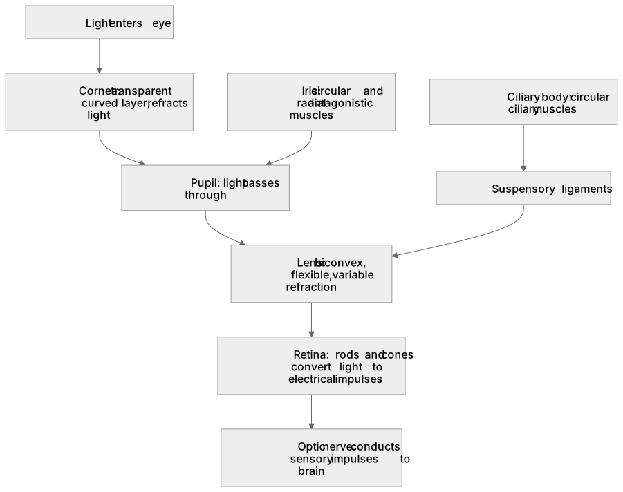
Key words
Say what each word means before you turn the card.
pupillary reflex
Try answering aloud or in your head before turning the card.
accommodation
Try answering aloud or in your head before turning the card.
eye structure
Try answering aloud or in your head before turning the card.
See every definition
- pupillary reflex
- Circular and radial antagonistic smooth muscles in the iris alter pupil diameter to regulate light entry during the pupillary reflex.
- accommodation
- The ciliary body contains circular ciliary muscles connected to the lens by suspensory ligaments, which control lens curvature during accommodation.
- eye structure
- The main structures of the eye include the cornea, iris, pupil, lens, retina, optic nerve, ciliary body and suspensory ligaments.
Label the diagram
Some boxes have lost their names. Pick the right name for each number.
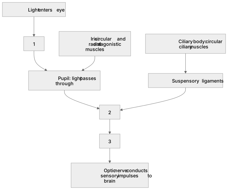
1Box 1
2Box 2
3Box 3
There is more to this lesson
The full lesson goes on to check your understanding and help you earn the marks. Create a free account to carry on, with your progress saved.
A week of Premium from your first lesson · Then the free plan continues · No card, no charge
Test your understanding of Coordination and control – the nervous system: explain how the main structures of the eye are related to their functions (to include: cornea, iris, pupil, lens, retina, optic nerve, ciliary body, suspensory ligaments) (content found only in the biology separate science qualification)
Active recall beats re-reading. Take an instant quiz now to test what you’ve learned and lock it into long-term memory.
A week of Premium from your first lesson · Then the free plan continues · No card, no charge